
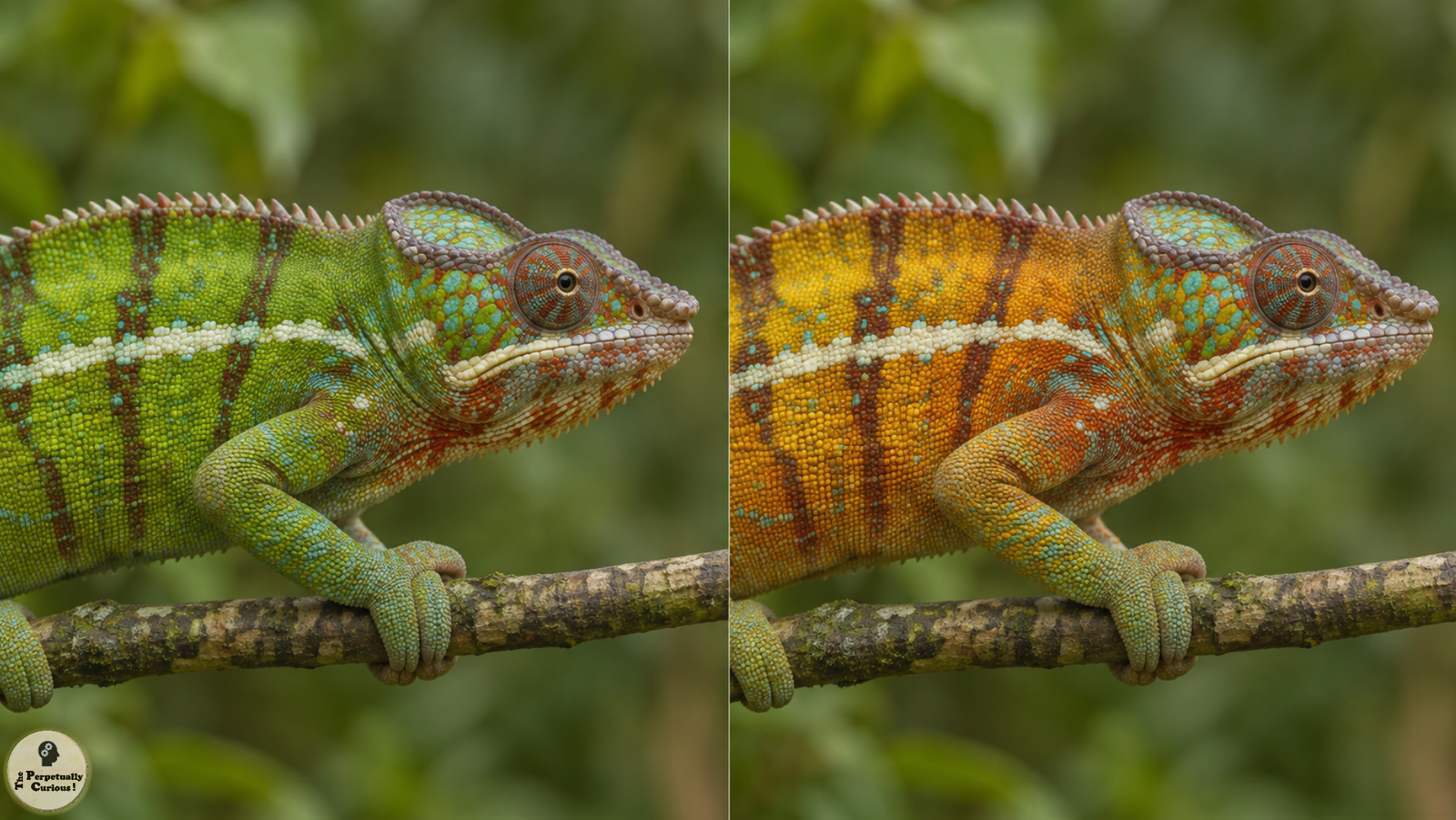
During an encounter with a rival, an adult male panther chameleon (Furcifer pardalis) can turn patches of green skin yellow or orange. The green can return as the display subsides. Beneath that visible shift is a change too small for the eye to see. The gaps between tiny crystals in the skin widen.[1]
Just beneath the skin surface are light-reflecting cells called iridophores. In the upper layer of these cells, tiny crystals of a substance called guanine form a repeating array. Light waves scattered by neighboring crystals can take slightly different paths. At some wavelengths the waves arrive in step and reinforce one another. The spacing of the crystals helps determine which wavelengths get that boost. A repeating structure that affects light in this way is called a photonic crystal.[1]
Reflection from the crystals is one step in producing the color of the patch. At rest, closely spaced crystals favor shorter wavelengths, toward the blue end of visible light. Yellow pigments in the skin selectively absorb some of the light. What reaches the eye has been shaped by both processes, which is how a reflector that favors blue can contribute to skin that looks green.[1][2]
What changes during a display
As the display begins, the gaps between the crystals widen. The array then favors longer wavelengths, changing the light available to interact with the pigments. A patch that looked green can now look yellow or orange. The material in the crystals is still guanine. Its arrangement has changed enough to shift the visible color.[1]
This is a form of structural color, in which the physical arrangement of a material helps determine its appearance. The shifting colors in peacock feathers offer another way to see that relationship. A feather can take on a different sheen as it tilts because the structures within it reflect different wavelengths at different angles. During the chameleon’s display, it is the spacing within the reflector that changes.[1][3]
Researchers traced the change by comparing electron microscope images of skin sampled while the animals were at rest and during a display. The display samples showed wider gaps between crystals of comparable size. In a separate test, they changed the conditions around skin samples in a way expected to bring the crystals closer together. The reflected light shifted toward shorter wavelengths. An optical model also reproduced much of the observed color shift by varying the spacing while keeping the crystal size fixed. The study left open how a living cell adjusts those gaps during a display.[1]
The same animal can also change how light or dark its skin looks. Dark pigment granules can spread through a cell or gather in a smaller area, changing how much light they absorb. Brightness can change alongside hue, the quality that distinguishes one color from another. On a displaying male, a red marking can grow brighter while a green patch shifts toward orange.[1][2]
The study reported changes in brightness in both sexes and across ages. The upper layer of iridophores was most developed in the adult males examined, and the vivid shift in hue described here was characteristic of their displays.[1]
As the display fades, the gaps narrow and the patch can return to green. A change in spacing that escapes the eye becomes visible in the color of the skin.[1]
Selected sources
- Teyssier J, Saenko SV, van der Marel D, and Milinkovitch MC. (2015). Photonic crystals cause active colour change in chameleons. Nature Communications, 6, 6368.
- Ligon RA and McCartney KL. (2016). Biochemical regulation of pigment motility in vertebrate chromatophores: a review of physiological color change mechanisms. Current Zoology, 62(3), 237–252.
- Zi J et al. (2003). Coloration strategies in peacock feathers. Proceedings of the National Academy of Sciences, 100(22), 12576–12578.