

Every heartbeat sends blood through a vast branching network, carrying oxygen, nutrients, hormones, medicines, and the chemical traces of life throughout the body. Yet when that circulation reaches the brain’s smallest vessels, the rules of passage change. The blood still nourishes the tissue, but it does not mingle freely with the delicate environment around neurons.
This selective frontier is known as the blood-brain barrier. It is one of the brain’s essential protections and one of medicine’s most persistent obstacles. The same system that limits the entry of toxins, pathogens, and disruptive fluctuations can also exclude a treatment designed to help. To understand that paradox, it helps to leave behind the image of an impenetrable wall and look instead at a living border that sorts, transports, senses, and adapts.
🧱 The barrier is built into the brain’s smallest vessels
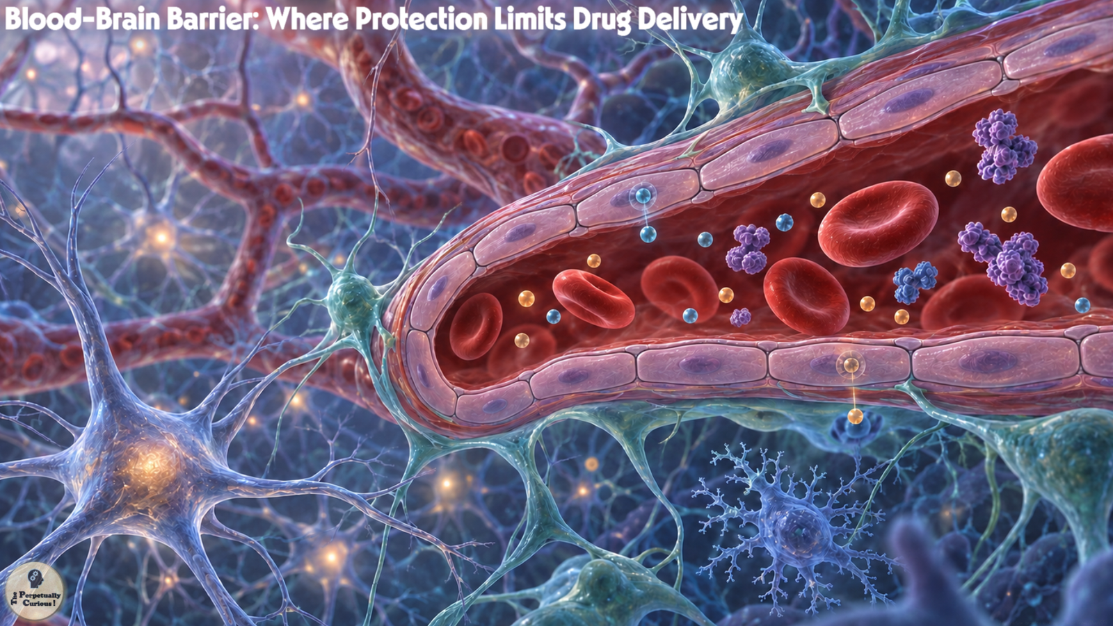
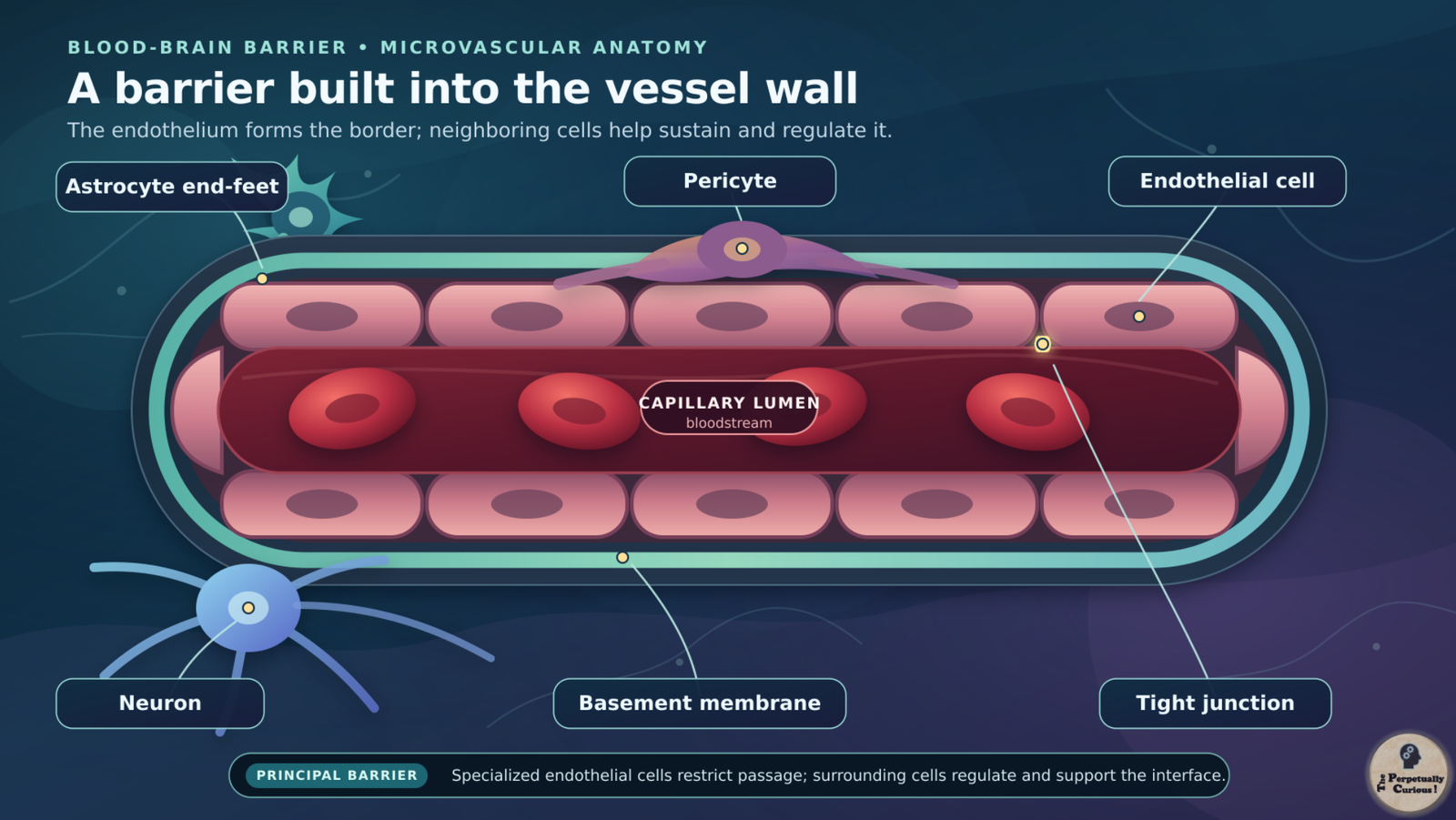
The blood-brain barrier is not a membrane wrapped around the brain. Most of it is formed by the endothelial cells that line microscopic blood vessels within the brain and spinal cord. Elsewhere in the body, gaps and active vesicular transport often allow substances to move comparatively freely between blood and surrounding tissue. Brain endothelial cells are different. They are joined by complex tight junctions and normally maintain unusually low levels of nonspecific transport through the cells.[1]
Those endothelial cells do not work alone. A basement membrane supports the vessel wall. Pericytes closely associate with the capillaries and help regulate barrier properties, vessel stability, and blood flow. Astrocyte end-feet surround much of the vessel surface and contribute signals that help maintain the local environment. Neurons, immune cells, and other glia also participate in the wider neurovascular unit, linking vascular behavior to the activity of neurons, glia, and networks.
The barrier is therefore better understood as a coordinated interface than as a single layer of biological brickwork. Its structure explains how passage is restricted, but its deeper importance lies in what it permits.
🚦 A border that sorts rather than seals
Some small gases, including oxygen and carbon dioxide, cross readily. Certain small lipid-soluble compounds can also diffuse through endothelial cell membranes, although size and fat solubility alone do not determine whether a substance will reach useful concentrations in the brain. Water moves through regulated pathways, while glucose, amino acids, ions, vitamins, and other necessities depend heavily on specialized channels and transport proteins. For a familiar point of comparison, consider caffeine and brain signaling.
Larger molecules may sometimes use receptor-mediated transport. Insulin and transferrin-related pathways, for example, allow selected cargo to be taken into endothelial cells and moved across them. In the opposite direction, efflux pumps can recognize many foreign compounds and send them back toward the blood. Enzymes within and near the barrier add another layer of chemical filtering.[1]
This selectivity is not perfect or identical everywhere. Small regions known as circumventricular organs have unusually permeable capillaries so that the brain can sample aspects of the blood or release hormones. The area postrema, for example, samples circulating signals involved in nausea and vomiting. These regions have their own surrounding boundaries and should not be imagined as unguarded holes into the rest of the brain. Immune surveillance also continues through regulated routes, especially at interfaces such as the meninges and choroid plexus.
The brain is therefore sheltered without being isolated. Signals from the body can still reach it through nerves, hormones, immune pathways, metabolites, and carefully controlled transport. Protection depends not on silence from the outside world, but on regulating how that world is heard.
🛡️ Why the brain needs this degree of control
Neural signaling depends on a precisely regulated chemical environment. Changes in ions, excitatory molecules, inflammatory mediators, or plasma proteins can disturb the electrical and metabolic conditions in which neurons operate. The barrier helps stabilize those conditions while allowing the continuous delivery of fuel and raw materials.
It also reduces exposure to many circulating toxins and microorganisms. This protection is substantial, but it is not absolute. Some pathogens can infect the central nervous system by crossing barrier cells, moving through infected immune cells, traveling along nerves, or entering through other anatomical interfaces. Some harmful chemicals cross because their properties resemble those of permitted molecules or because transport systems carry them.
That combination of strength and selectivity is central to the barrier’s success. It is also why a medicine cannot be judged only by what it does to a cell in a laboratory dish. Before it can act inside the brain, it must first arrive at the right place in a usable form.
💊 When protection becomes a treatment bottleneck
A medicine circulating in the blood encounters several questions at once. Can enough of it remain unbound and stable? Can it cross or use a transporter? Will an efflux pump remove it? Can it spread through brain tissue after crossing? Will it reach the intended cells at an effective concentration without causing unacceptable effects elsewhere in the body?
Small molecules sometimes pass, but many do not. Large proteins, antibodies, enzymes, and nucleic-acid medicines generally face even greater difficulty because they do not diffuse freely through the barrier. Increasing a systemic dose may raise exposure in the rest of the body without producing a proportional increase at the neural target. The barrier can therefore turn an otherwise promising compound into a delivery problem.[2]
Even successful entry is not the end of the journey. A drug may enter endothelial cells but become trapped or broken down within them. It may reach cerebrospinal fluid without penetrating deeply into brain tissue. It may accumulate in one region while missing another. For brain treatment, “crossing the barrier” is not a single finish line. It is a chain of biological distances.
🧩 The barrier changes across disease, place, and time
The blood-brain barrier is dynamic. Its properties can vary among brain regions and may change with development, aging, inflammation, infection, stroke, traumatic injury, epilepsy, multiple sclerosis, neurodegeneration, and cancer. These changes can involve tight junctions, transporters, immune-cell traffic, pericytes, astrocytes, or the basement membrane.
Barrier disruption does not necessarily make treatment easier. A leak may be patchy, temporary, or concentrated in the wrong region. It can admit inflammatory proteins and disturb tissue while still failing to deliver enough medicine to the cells that need it. In brain tumors, vessels near an abnormal core may be relatively permeable, while infiltrating tumor cells at the margins can remain protected by vessels with stronger barrier properties. The resulting blood-tumor barrier is heterogeneous rather than uniformly open.
This matters because a disease can produce two opposing problems at once. The brain may lose some of the protection that preserves its internal balance, yet remain insufficiently accessible to therapy.
🧬 Neurological disorders reveal the central paradox
Many neurological disorders affect widely distributed cells or networks. A treatment for a focal lesion may need to reach one region, while a treatment for a neurodegenerative or inherited metabolic disorder may need broad access across the brain. Molecular size, disease location, the condition of the barrier, and the biology of the target all influence what kind of delivery is plausible.
The challenge is especially visible in disorders involving protein misfolding. A molecule intended to alter amyloid, tau, alpha-synuclein, or another disease-related process may show activity in experimental systems but still face limited and uneven access in the human brain. Brain cancers present a different version of the same problem because the therapeutic target can extend beyond the visibly abnormal vessels of the tumor core.
Yet the barrier is only one part of these diseases. Neuronal vulnerability, genetics, inflammation, timing, target selection, and the stage of illness also shape treatment outcomes. Better delivery cannot rescue a medicine aimed at the wrong mechanism. It can, however, determine whether a biologically sound treatment ever has the chance to act.
That distinction leads to the practical question at the heart of current research: if the guarded border cannot simply be removed, how might medicine negotiate passage?
🚪 There is no single route into the brain
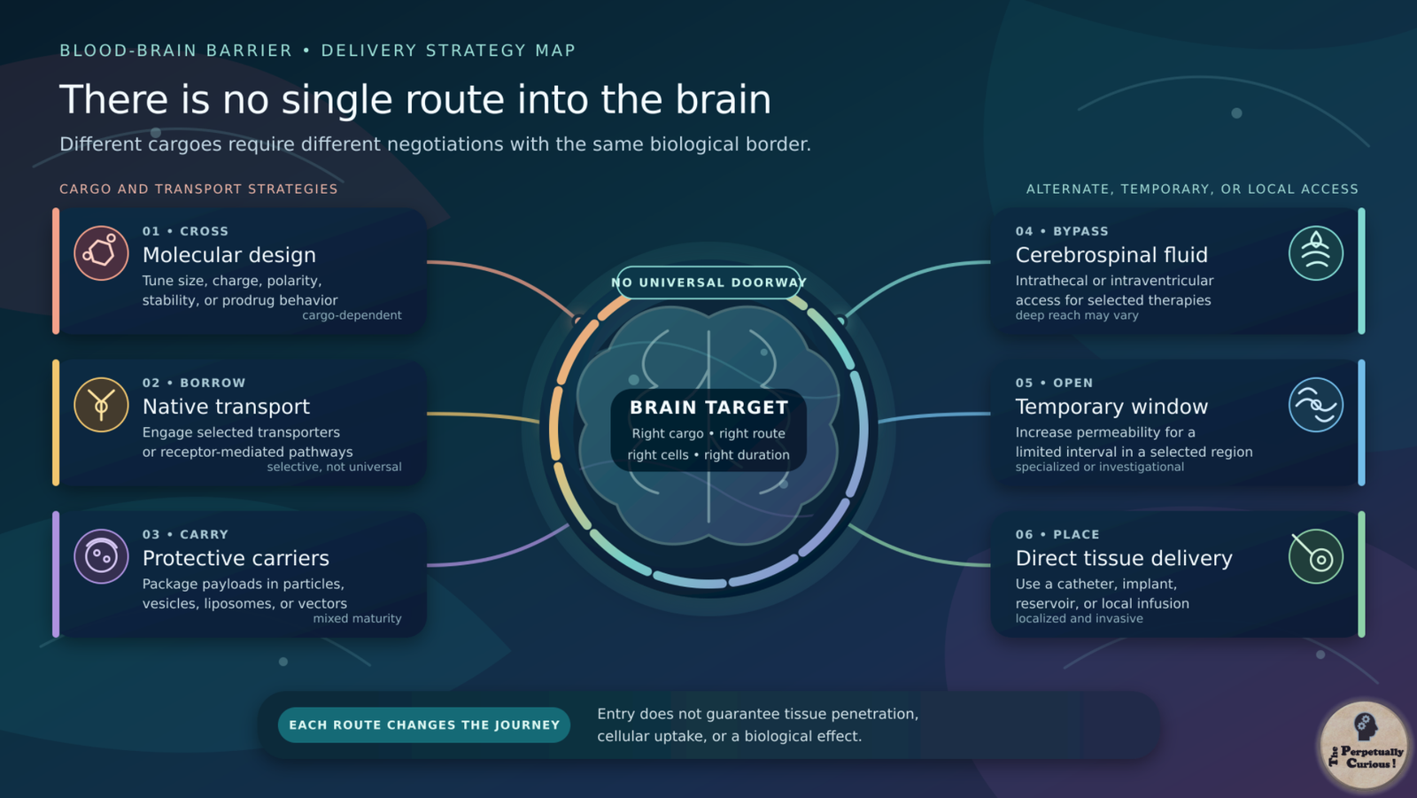
Strategies for brain delivery fall into several broad families. A medicine can be designed to cross the barrier, attached to a molecular shuttle, carried within a delivery system, introduced into cerebrospinal fluid, delivered directly into tissue, or paired with a method that opens a localized and temporary window. Each approach solves one problem while creating others.[2]
The best route depends on the cargo and destination. A small molecule intended for widespread daily exposure presents a different challenge from an enzyme, an antibody, a gene therapy, or a drug meant for one tumor site. The methods are therefore not competing versions of one universal doorway. They are different negotiations with the same biological border. The first negotiation begins with the medicine itself.
🧪 Designing a medicine that can cross
Medicinal chemists can adjust molecular size, charge, polarity, fat solubility, stability, and affinity for transport or efflux systems. A compound must be soluble enough to move through the body, stable enough to survive, and selective enough to avoid unwanted targets. Making it more lipid-soluble may improve passive entry in some cases, but excessive lipid solubility can reduce solubility, increase nonspecific binding, alter metabolism, or create effects in other tissues.
Prodrugs offer another route. An active medicine can be temporarily modified into a form that crosses more readily, then converted back after entry. The familiar example is levodopa. Dopamine itself does not cross the blood-brain barrier effectively, but levodopa resembles a large neutral amino acid and uses an amino-acid transporter to enter the brain, where it can be converted to dopamine.
This approach is powerful when the chemistry and biology align. It is less useful for very large or fragile cargo, and even small molecules can be expelled by efflux pumps. Molecular design can persuade the gate, but it cannot make every passenger resemble one the brain already expects. When chemistry alone cannot make the cargo suitable for passage, the next option is to borrow routes already operating at the barrier.
🔑 Borrowing the brain’s transport machinery
This second strategy uses the barrier’s own carriers and receptors. A therapeutic cargo can be engineered to engage a transporter or attached to a molecule that binds a receptor capable of moving material through endothelial cells. Transferrin-receptor and insulin-receptor pathways are among the most studied examples.
This idea is sometimes described as a molecular shuttle or Trojan horse, but successful transport requires more than strong binding. Cargo must enter the appropriate cellular route, avoid degradation, disengage on the brain side, and retain its biological activity. Binding too tightly can sometimes trap a construct within the endothelial cell or alter receptor handling. Receptors may also be present in other organs, creating distribution and safety concerns.
Transporter-based medicines already demonstrate that native routes can be used clinically. Engineered receptor shuttles for larger biologic medicines are advancing through preclinical and clinical development, but they are not yet a universal platform for brain delivery. The promise lies in selectivity. The difficulty lies in preserving it through every step of the journey.
🎯 Packaging cargo without losing the destination
When therapeutic cargo cannot travel alone, a protective carrier offers another negotiation with the border. Liposomes, polymer particles, lipid nanoparticles, extracellular vesicles, and viral vectors can protect a payload and influence where it travels. Their surfaces may be modified with molecules intended to improve circulation time, bind a target, or engage a transport pathway. Some systems can also control when or where a payload is released.
These carriers are not invisible boats that automatically sail into the brain. Many are cleared by the liver, spleen, or immune system. Some accumulate mainly in blood vessels rather than crossing into neural tissue. Evidence that a carrier appears in a whole-brain sample does not by itself prove that an active payload reached the intended cells. Size, surface chemistry, dose, manufacturing consistency, immune response, and the condition of the barrier all matter.
Several carrier and vector approaches have entered clinical use or clinical testing for particular diseases and routes, while many brain-targeted nanoparticle concepts remain preclinical. The field is broad because “delivery system” describes a toolbox, not one method with one level of evidence.
🌊 Entering through the brain’s fluid spaces
Not every strategy asks a medicine to cross from the bloodstream. Some treatments bypass the vascular barrier by entering the cerebrospinal fluid that surrounds the brain and spinal cord. Intrathecal administration introduces a medicine into the spinal fluid, while intraventricular administration places it within the brain’s ventricles, sometimes through an implanted reservoir. These routes are established for selected anesthetics, anti-infective medicines, cancer treatments, antisense medicines, enzyme therapies, and gene therapies.
Bypassing the blood-brain barrier does not guarantee even delivery throughout the brain. Cerebrospinal fluid circulates and clears substances, while movement from fluid spaces into deep tissue can remain limited by diffusion, binding, cellular uptake, and anatomy. A treatment may reach the spinal cord or surfaces near the fluid more readily than a distant target within the brain’s parenchyma. The wider journey of cerebrospinal fluid therefore becomes part of the delivery problem.
These routes can permit large molecules to reach the central nervous system, but they are more invasive than ordinary oral or intravenous dosing and can bring procedure-related risks. They bypass one border while encountering the geography beyond it.
Intranasal delivery is also being studied as a less invasive route. Molecules placed in the nasal cavity may follow pathways associated with the olfactory and trigeminal nerves, enter the bloodstream through the nasal lining, be swallowed, or be removed by mucociliary clearance. Direct nose-to-brain delivery has been demonstrated in experimental settings, but the amount and distribution achieved in humans can be difficult to establish. It is therefore better regarded as a route with specific possibilities and limitations than as an open shortcut around the barrier.
🔓 Opening a temporary window
When molecular redesign and alternate routes are not enough, some methods temporarily increase barrier permeability. Hyperosmotic solutions delivered through specialized arterial procedures can cause endothelial cells to shrink and loosen junctional restriction for a limited period. This approach has been used in selected clinical settings, particularly for regional delivery in neuro-oncology, but it is invasive and not precisely confined at the microscopic level.
Focused ultrasound offers a more localized experimental strategy. Low-intensity ultrasound aimed at a selected brain region can interact with circulating microbubbles, producing mechanical effects that temporarily increase permeability in nearby vessels. Magnetic resonance imaging and acoustic monitoring may be used to guide the target and assess the response.
This low-intensity barrier-opening approach should not be confused with higher-intensity focused ultrasound procedures that intentionally heat and ablate selected brain tissue. Some ablation procedures are approved for particular movement disorders, while focused-ultrasound opening for drug delivery remains a separate investigational use.
A small Alzheimer’s disease study combined aducanumab infusions with focused ultrasound in three participants, using ultrasound to temporarily increase blood-brain barrier permeability.[3] This provided an early demonstration of the approach, while ultrasound-assisted drug delivery remains under clinical investigation. Further research must establish how consistently medicines reach their intended targets, assess tissue safety over repeated procedures, and determine whether the approach improves clinical outcomes.
The attraction of a temporary window is clear. It may allow treatment into one region without changing the medicine itself. The responsibility is equally clear because opening a protective border requires control over where, how much, and for how long.
🧭 Going directly to the tissue
A different answer is to shorten the journey. For a localized target, medicine can sometimes be placed directly into or beside brain tissue. Convection-enhanced delivery uses catheters and a pressure gradient to distribute an infused treatment through a selected region. Other approaches include local injections, biodegradable implants, drug-releasing wafers, and implanted reservoirs.
Direct delivery can achieve high local exposure while reducing the need for a drug to cross from blood.[4] Its limitations arise from the same precision that makes it attractive. Catheter position, tissue structure, backflow, pressure, and clearance can produce uneven distribution. Surgery and implanted hardware add risks, and a local route may not suit a disease spread across many regions.
The method therefore changes the scale of the problem. Instead of asking whether a drug can cross every vessel, it asks whether a carefully placed source can reach enough of the surrounding tissue.
⚖️ Every shortcut creates a new boundary
No delivery strategy simply defeats the blood-brain barrier. Chemical redesign can affect potency and off-target behavior. Molecular shuttles can encounter receptor saturation or trapping. Carriers can be cleared before reaching the brain. Fluid-space delivery may not penetrate deeply. Temporary opening can admit unintended blood components. Direct infusion trades a vascular barrier for an invasive procedure and a distribution challenge.
These are not reasons to dismiss the methods. They explain why progress often comes through combinations. A carrier may be paired with a receptor shuttle. A systemic medicine may be combined with focused ultrasound. A local infusion may use imaging to map distribution. The useful question is not only “Did the treatment enter the brain?” but “Did the active treatment reach the right cells, at the right concentration, for the right duration, with an acceptable balance of risk?”
Answering that question requires researchers to measure passage rather than merely assume it.
🔬 Measuring what crossed is part of the challenge
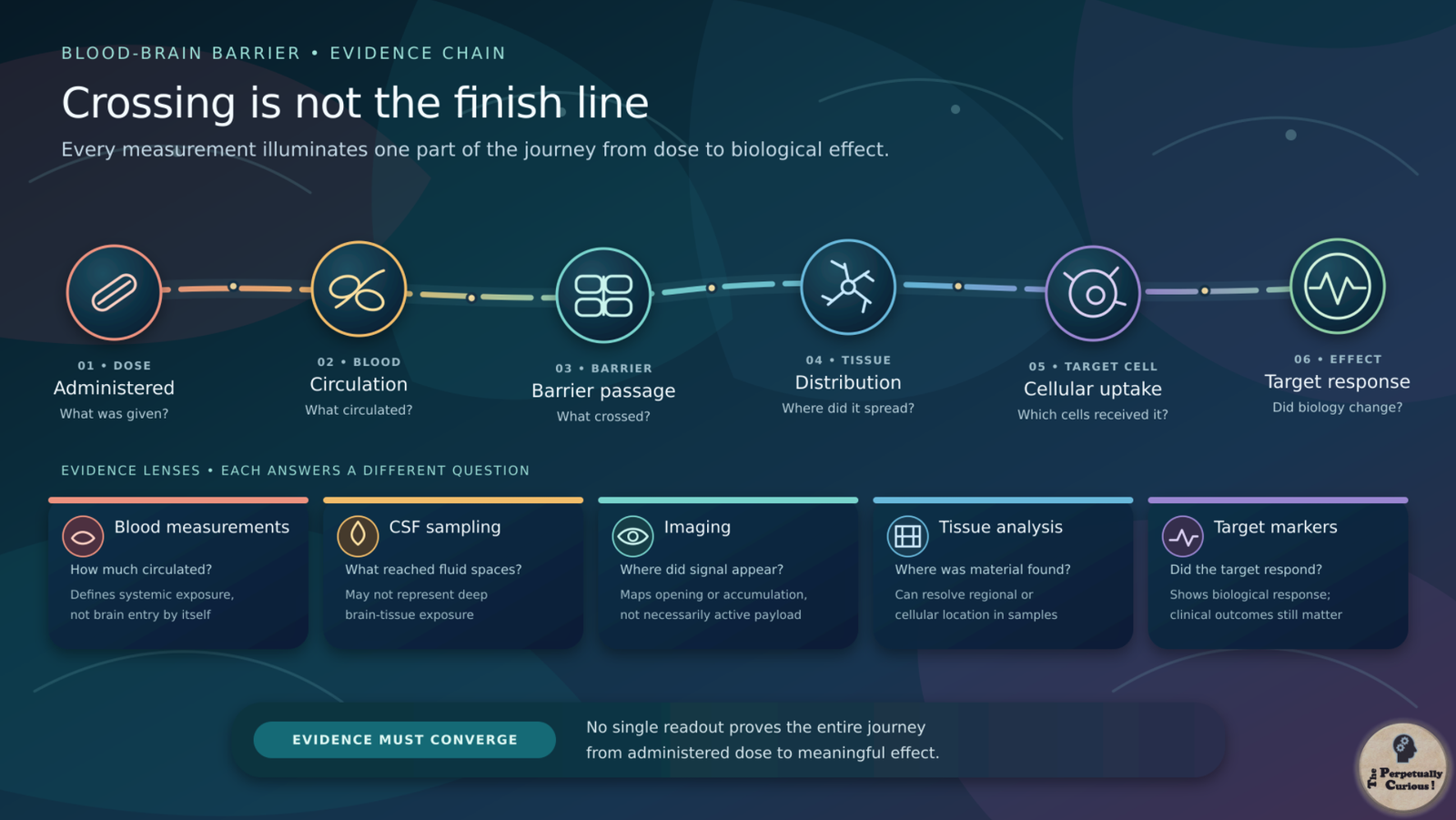
Brain delivery can be studied through imaging tracers, magnetic resonance contrast, cerebrospinal fluid sampling, blood measurements, surgical tissue samples, and biomarkers showing that a target has been affected. Each method reveals only part of the picture. A concentration in blood says little about free drug inside brain tissue. A concentration in cerebrospinal fluid may not represent deep parenchymal exposure. A signal in a whole-brain sample may include medicine still trapped within blood vessels.[5]
Researchers therefore combine measurements whenever possible. Imaging can show where a barrier has opened or where a labeled molecule accumulates. Tissue analysis can reveal cellular location. Pharmacodynamic markers can indicate whether the treatment changed its intended target. Clinical outcomes remain essential because improved delivery is valuable only when it contributes to a meaningful and sufficiently safe effect.
This measurement problem is one reason dramatic laboratory results can be difficult to translate. The final distance is not from blood to brain in the abstract. It is from a dose to a verified biological effect.
🌅 The future may favor controlled passage over an open gate
The most promising future is unlikely to be a permanently weakened barrier. It is more likely to involve controlled passage shaped by the molecule, disease, target region, and individual barrier state. Better human cell models, organ-on-chip systems, molecular imaging, computational prediction, and tissue-level measurements are helping researchers study those differences before and during clinical trials.
Some therapies may be designed to cross quietly through native transport systems. Others may arrive through cerebrospinal fluid, a local device, or a temporary opening measured in real time. Still others may work from outside the brain by changing immune, metabolic, or neural signals that the brain already receives. Progress may come not from discovering one master key, but from learning which doorway belongs to which treatment.
The blood-brain barrier embodies a difficult form of biological wisdom. The brain must remain connected to the body without being exposed to every fluctuation within it. Medicine faces the same task in reverse: to enter without undoing the protection that made entry difficult. At this guarded border, the future of treatment depends not on breaking the gate, but on understanding the intelligence of its design.
Pass this article along to someone curious and let the learning travel.
💡 Did You Know?
🎨 In the late nineteenth century, Paul Ehrlich observed that dyes injected into the bloodstream stained many organs but largely spared the brain. In the early twentieth century, Edwin Goldmann obtained a contrasting pattern after introducing dye into cerebrospinal fluid. These observations helped reveal that blood, brain tissue, and cerebrospinal fluid are separated by selective interfaces, although the modern barrier concept emerged gradually.
🫧 A transport protein called MFSD2A performs two very different barrier tasks. It helps carry certain docosahexaenoic acid, or DHA, containing lipids into the brain and also helps suppress nonspecific vesicle traffic through brain endothelial cells.
⚡ When a brain region becomes more active, local blood flow can rise through neurovascular coupling. Under ordinary conditions, greater flow delivers more oxygen and glucose without requiring the blood-brain barrier to become broadly leaky.
🌍 Barrier transport is not identical across species, ages, diseases, or brain regions. This variation is one reason a drug that enters the brain in a laboratory model may behave differently in a human clinical study.
🔋 Brain endothelial cells use energy-dependent transporters to move selected substances and expel others. Passage therefore depends on active cellular work as well as on the physical tightness of the vessel wall.
👶 The developing blood-brain barrier is not simply absent before birth or in early life. Barrier features arise during development and continue to mature. Later in life, transport, vascular support, and permeability may also change, especially in some diseases, but aging does not produce one uniform barrier state across every person or brain region.
What is the blood-brain barrier in simple terms?
The blood-brain barrier is a selective interface formed mainly by specialized cells lining the brain’s smallest blood vessels. It regulates movement between the bloodstream and neural tissue, admits essential substances through controlled routes, and limits many potentially disruptive molecules.
Does the blood-brain barrier block all medicines?
No. Many medicines that act on the brain can cross by passive diffusion or use transport systems. Others reach only low concentrations, are removed by efflux pumps, or cannot cross effectively because of their size, charge, polarity, or biological structure.
Can large medicines such as antibodies or enzymes reach the brain?
Only limited amounts of many large biologic medicines cross an intact barrier on their own. Researchers may engineer them to use receptor-mediated transport, deliver them into cerebrospinal fluid, place them locally, or combine them with a temporary opening method. The appropriate approach depends on the disease, target, and cargo.
Does a damaged blood-brain barrier make treatment easier?
Not reliably. Damage can be patchy and may expose brain tissue to inflammatory or toxic blood components without delivering enough medicine to the intended cells. In tumors and other disorders, permeability can differ across regions and change over time.
What is the blood-tumor barrier?
The blood-tumor barrier is the altered vascular interface found within and around many brain tumors. It is not uniformly open. Vessels in an abnormal tumor core may be relatively permeable, while infiltrating tumor cells at the margins can remain behind vessels with stronger barrier properties. This unevenness can make drug distribution difficult to predict.
Is the blood-brain barrier the same as the blood-cerebrospinal fluid barrier?
No. The blood-brain barrier is formed mainly by endothelial cells in brain microvessels. The blood-cerebrospinal fluid barrier is formed largely by epithelial cells of the choroid plexus. The two interfaces are related, but they have different structures and transport properties.
Do intrathecal medicines reach the entire brain?
Intrathecal administration places medicine into cerebrospinal fluid and can distribute some treatments broadly through the central nervous system. However, entry from that fluid into deep or distant brain tissue may remain uneven, and the pattern depends on the molecule, dose, anatomy, clearance, and target.
How can the gut, hormones, or immune system influence the brain if the barrier exists?
The body can communicate with the brain through several regulated routes. Nerves carry electrical signals, selected hormones and metabolites use transport pathways or act at specialized interfaces, and immune signals can alter vascular and neural activity. This layered gut-brain communication does not require unrestricted movement of material from the intestine into the brain.
Can focused ultrasound open the blood-brain barrier?
Low-intensity focused ultrasound used with circulating microbubbles can temporarily increase permeability in a selected region. Human studies have shown that this can be done under controlled research conditions, but its use for routine drug delivery remains investigational. Safety, dosing, repeatability, and clinical benefit continue to be studied.
Why do researchers use several models to study the blood-brain barrier?
No single model reproduces every aspect of the human barrier. Cell cultures can isolate molecular mechanisms. Organ-on-chip systems can reproduce selected features of flow and interactions among barrier cells. Animal models reveal whole-body distribution and responses, while human imaging, cerebrospinal fluid, and tissue studies show how delivery behaves clinically. Confidence grows when findings converge across complementary methods.
Can the blood-brain barrier itself contribute to neurological disease?
Barrier dysfunction may contribute to inflammation, edema, altered transport, or exposure to harmful blood components in several disorders. Its role varies by condition, region, and disease stage, and it is usually one part of a wider biological process rather than a single cause.
🌸 Sharing the wonder across a guarded threshold
We kindly invite you to share and spread the word. If this journey across the brain’s guarded border clarified why protection and treatment can become entwined, please pass it along to friends, colleagues, and fellow curious minds. Your support in helping thoughtful science reach a wider audience is deeply appreciated.
Some borders protect by remaining closed. The most remarkable ones teach us how life decides what may pass.
📚 Educational context
This article presents scientific information about the blood-brain barrier and methods studied or used to deliver medicines to the brain for educational purposes. It does not provide medical advice, diagnosis, or treatment guidance. Barrier integrity, drug access, and treatment response can vary by condition and individual circumstances. Questions about neurological symptoms, medicines, or treatment should be discussed with a qualified healthcare professional.
Selected sources
- Daneman R, Prat A. (2015). The blood-brain barrier. Cold Spring Harbor Perspectives in Biology, 7(1), a020412. [PMID: 25561720]
- Pandit R, Chen L, Götz J. (2020). The blood-brain barrier: Physiology and strategies for drug delivery. Advanced Drug Delivery Reviews, 165-166, 1-14. [PMID: 31790711]
- Rezai AR, D’Haese PF, Finomore V, et al. (2024). Ultrasound Blood-Brain Barrier Opening and Aducanumab in Alzheimer’s Disease. New England Journal of Medicine, 390(1), 55-62. [PMID: 38169490]
- Somayaji MR. (2016). Perspectives on Development and Regulation of Therapeutic Products for CED-Based Therapy of Neurodegenerative Diseases. Current Pharmaceutical Biotechnology, 17(6), 495-512. [PMID: 26517332]
- Hammarlund-Udenaes M, Fridén M, Syvänen S, et al. (2008). On the rate and extent of drug delivery to the brain. Pharmaceutical Research, 25(8), 1737-1750. [PMID: 18058202]
“The Brain’s Guarded Border: How the Blood-Brain Barrier Protects Us and Complicates Treatment.” The Perpetually Curious!, September 2026.
https://www.theperpetuallycurious.org/articles/blood-brain-barrier/Continue Exploring
For the curious mind: want to explore this theme through a few questions? Visit the Classic Quiz Studio, nestled under Explorations opens in a new tab ,and see where a few well-placed questions lead: Biology & Health Quizzesopens in a new tab
For what comes next
Site Updates gathers occasional notes, new additions, and ways to keep in touch. RSS brings new publications to your feed reader in chronological order.
Choose the path that speaks to you, and let the next curiosity find you wherever your wondering takes you.