

A mole can be so familiar that it barely registers: a small mark on a shoulder, a raised spot near the hairline, or a tiny feature that seems to have occupied the same patch of skin for years. Yet beneath that quiet surface is an intriguing biological event. A small population of melanocytic cells has expanded locally, creating a distinct cellular community within the surrounding skin.
Moles, medically called melanocytic nevi, are usually benign proliferations of melanocytic cells. They are therefore more than areas where the skin happens to contain extra pigment. Their visible color may draw our attention, but underneath is a story of cell growth, development, genetic change, tissue architecture, and biological restraint.
That turns an ordinary spot into a deeper question. Why does one small population of cells begin following a different path from the melanocytes around it? And once that growth begins, why does an ordinary mole usually reach a boundary and stop?

A small island of cells rests in its place, carrying its own quiet history beneath the surface.
Light moves across it without hurry, revealing only what the skin chooses to show.
🔬 A mole begins with cells, not simply with pigment
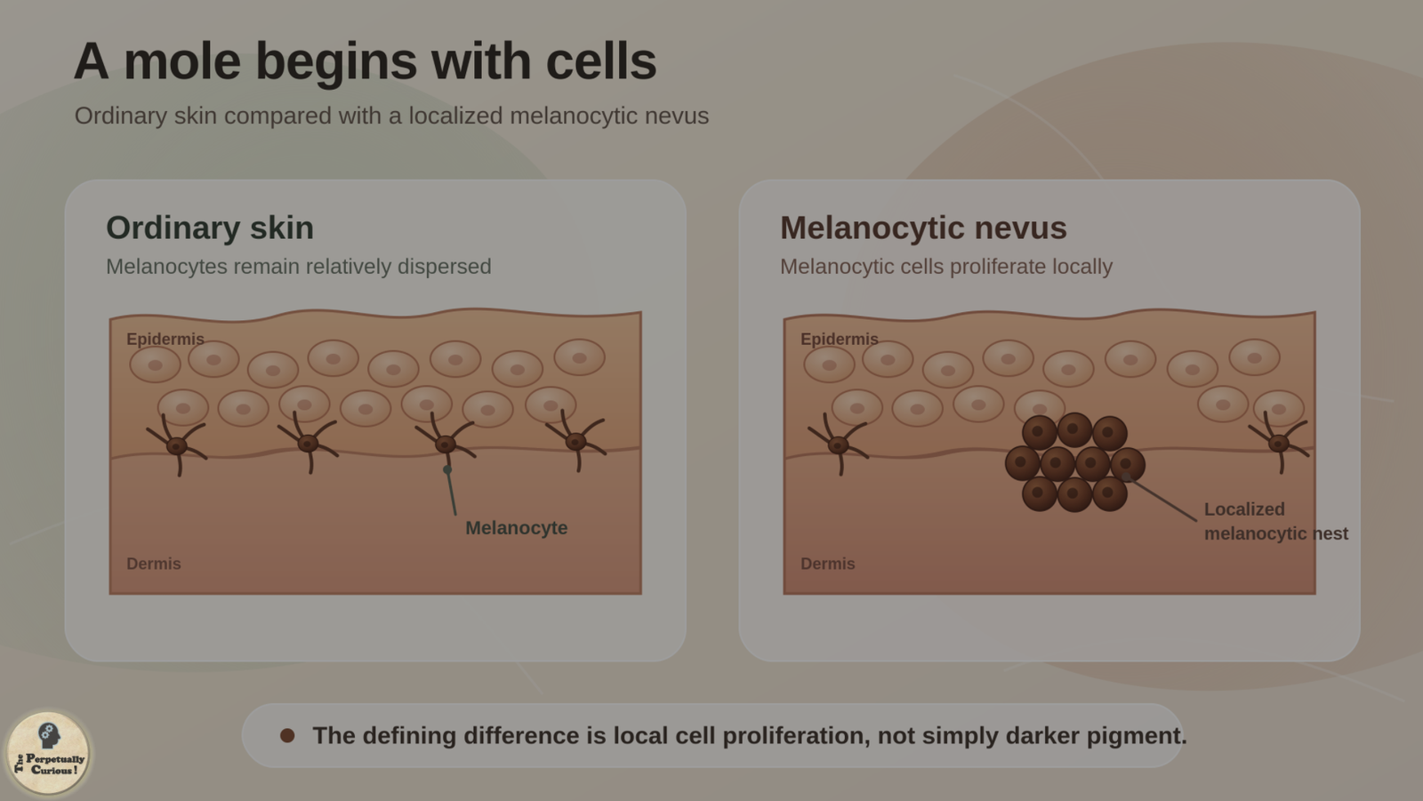
Melanocytes are specialized cells that produce melanin, a family of pigments that contributes to the coloration of skin, hair, and eyes. In ordinary epidermis, melanocytes generally occur as individual cells among their neighbors rather than as the localized cellular nests characteristic of many melanocytic nevi.
In a nevus, melanocytic cells proliferate locally and commonly organize into small groups or nests within the skin. The visible result can vary considerably because appearance reflects not only pigment, but also where the nevus cells lie and how they are arranged.
This distinction is important. A mole is not simply an unusually dark piece of otherwise ordinary skin. Pigment helps make many moles visible, but the defining event is the localized proliferation of melanocytic cells.
Once that cellular origin is clear, the next question moves beneath appearance and into history. What caused this particular population of cells to follow a different trajectory from its neighbors?
🧬 A tiny genetic change can start a much larger cellular story
Many acquired moles contain somatic genetic changes, meaning alterations that arise in particular cells during life rather than being inherited in every cell of the body. One of the best studied involves BRAF, a gene in a signaling pathway that helps regulate cell growth. Activating BRAF variants, particularly BRAF V600E, occur frequently in common acquired melanocytic nevi.
Not every nevus follows the same molecular route. Congenital melanocytic nevi begin during development before birth and often show a different genetic pattern. Postzygotic NRAS alterations are especially important in multiple and larger congenital melanocytic nevi, although congenital lesions are themselves biologically diverse.
The localized nature of these changes explains something easy to overlook. A genetic alteration can arise after fertilization in one developing cell lineage and then remain confined to only part of the body. This patchwork condition is called somatic mosaicism. Multiple congenital melanocytic nevi associated with postzygotic NRAS variants provide a particularly clear example.
A mole can therefore preserve a tiny patch of cellular history within otherwise ordinary skin.
Yet a local genetic event is only the beginning. Which nevi become visible, how many appear, and where they develop also reflect the wider biological setting in which those cells live.
☀️ Where sunlight enters the story
The relationship between sunlight and skin includes many distinct biological responses, and melanocytic nevi are part of that wider landscape. Studies in children have found associations between sun exposure and nevus development, while the broader biology makes clear that ultraviolet exposure is not required for every nevus to arise.
Congenital nevi begin before ordinary postnatal ultraviolet exposure, and BRAF V600E has also been detected in acquired nevi from relatively protected sites. Sunlight is therefore better understood as one contributor to some acquired-nevus patterns than as a universal initiating explanation.
The broader pattern emerges from several layers: melanocyte biology, local genetic events, inherited predisposition, developmental timing, environmental exposure, and local tissue context.
Those influences do not all act at the same stage of life. That is one reason the map of moles across the skin is not necessarily complete at birth.
🕰️ Why moles appear at different chapters of life
Some melanocytic nevi are present at birth or become apparent soon afterward because their developmental story began before birth. Many others are acquired later. Nevus counts rise markedly through childhood and adolescence, and population studies indicate that counts generally increase into early or middle adulthood before tending to decline later in life.
A mole is also not necessarily a mark that appears once and remains visually identical forever. Longitudinal studies show that many nevi remain stable while others change, shrink, become less conspicuous, or disappear. The natural history of nevi therefore includes both persistence and change rather than one universal life course.
People also differ greatly in the number of moles they develop. Genetic variation contributes substantially to nevus count, while age, pigmentation traits, sun exposure, and other biological influences help shape the visible pattern.
The result is less like a set of dots stamped onto the skin at one moment and more like a changing biological map. Some marks arrive early, some emerge later, many settle into prolonged stability, and some slowly recede.
That life history leads directly to the most intriguing question in the article. If a mole can begin growing and then settle into stability, what restrains the cells after that initial proliferation?
🛑 The deeper puzzle is not why a mole grows, but why it stops
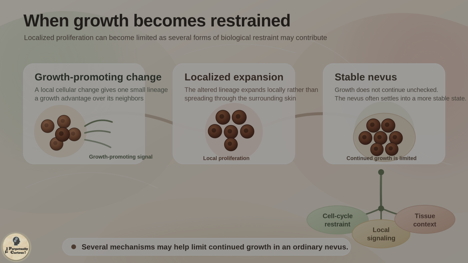
An activating growth signal might sound as though it should make cells continue dividing indefinitely. Ordinary melanocytic nevi show why living tissues are more complicated than that expectation.
One influential explanation is oncogene-induced senescence, in which an oncogenic growth signal becomes associated with a strong arrest of proliferation. Classic experiments showed that sustained BRAF V600E expression in human melanocytes can produce growth arrest accompanied by senescence-associated features.
Later research complicated the idea of one permanent switch. BRAF V600E-induced arrest in primary human melanocytes has been shown experimentally to be both conditional and reversible under particular cellular conditions. These findings caution against assuming that every nevus cell occupies one identical, irreversible state.
The central biological observation remains striking. A common nevus can harbor a strong growth-promoting alteration and still settle into remarkably limited growth. The molecular signal that helps begin the lesion does not, by itself, dictate indefinite expansion.
That restraint also clarifies an important distinction. A genetic change capable of promoting proliferation is not, by itself, the same thing as malignant growth.
⚖️ A benign growth is not the same thing as uncontrolled growth
The word mutation often carries an alarming association because mutations are frequently discussed in relation to cancer. In living tissues, however, genetic changes arise in cells throughout life, and their consequences depend on which genes are altered, what additional changes occur, and whether growth-control mechanisms remain effective.
A melanocytic nevus can contain an oncogenic BRAF alteration while remaining benign. BRAF activation is frequent in ordinary acquired nevi, yet that alteration alone is insufficient to produce melanoma.
Melanoma can arise in association with a pre-existing nevus, but it can also arise independently. Progression toward melanoma involves additional biological changes beyond the events that can establish a benign nevus.
The important contrast is therefore not simply mutation versus no mutation. It is restrained versus progressively dysregulated growth.
Once that distinction is clear, the story can move back from molecular behavior to something visible in everyday life: why one mole may look quite different from another.
🎨 Every mole carries a slightly different history
Two moles on the same person may differ in color, height, texture, and prominence. Part of that variation reflects where nevus cells are arranged within the architecture of the skin.
Junctional nevi are typically flatter because their nevus cells are concentrated near the epidermal-dermal junction, while intradermal nevi are commonly elevated because their cells reside within the dermis. Compound nevi contain components in both regions and can show intermediate appearances. These categories describe architecture rather than a mandatory step-by-step life cycle through which every mole must pass.
Nevus cells can also change in microscopic appearance with depth. In many ordinary nevi, cells deeper in the dermis become progressively smaller and show other changes collectively described as maturation. It is a common histologic pattern rather than a feature that every melanocytic lesion displays in exactly the same way.
Visible marks on skin can therefore resemble one another while arising from completely different biological processes. palm lines, for example, arise from developmental anatomy and the mechanics of the hand, while a melanocytic nevus reflects a localized proliferation of melanocytic cells. Similar-looking surface features do not necessarily share an origin.
Pigment adds another layer. A mole does not have to be dark brown to be a melanocytic nevus. Its apparent color reflects the amount and distribution of melanin, the depth and arrangement of nevus cells, and the pigmentation of surrounding skin.
Each mole is therefore less like a drop of color placed onto the body and more like a small biological history written into it.
🌿 Small marks, quiet boundaries
There is something striking about an ordinary mole when it is viewed at cellular scale.
A small population of cells begins following a distinct trajectory. It expands locally. A visible mark emerges. Yet in most common nevi, that expansion encounters biological restraints and settles into long-term stability.
The familiar spot on the skin therefore sits at the meeting point of several larger ideas: development, pigmentation, somatic change, environmental influence, and the regulation of cellular growth.
Moles do not need to perform a special adaptive function to be biologically revealing. Their value as a curiosity lies in what they expose about living tissue. A cellular lineage can acquire its own local history. A growth signal can begin a process without dictating its ultimate outcome. Biological change can coexist with biological restraint.
What appears from the outside as a tiny mark may therefore preserve an unexpectedly intricate balance between change and stability, proliferation and restraint.
Pass this article along to someone curious and let the learning travel.
💡 Did You Know
🎨 Not every patch of color on the skin is produced in the same way. A mole contains a localized proliferation of melanocytic cells, while henna stains skin when lawsone from the plant interacts with keratin in the outer layers of the skin. The two can both create visible color while operating through fundamentally different biology and chemistry.
🪶 Some hairy nevi appear to actively encourage hair growth rather than merely leaving nearby follicles undisturbed. A 2023 study found elevated osteopontin signaling from senescent melanocytes in hairy nevi; osteopontin promoted hair growth in experimental models and stimulated human hair follicles. The finding should not be assumed to explain every hair-bearing mole.
🔬 In many ordinary nevi, melanocytic cells become smaller and change in other ways as they extend deeper into the dermis. Pathologists refer to this common pattern as maturation, although it is not displayed identically by every melanocytic lesion.
🌙 Some moles become less conspicuous with age. Longitudinal studies show that individual nevi can remain stable, change, shrink, or disappear, illustrating that a mole is not necessarily a permanently frozen structure.
👶 A congenital melanocytic nevus is not necessarily inherited from a parent. Many congenital nevi arise through postzygotic somatic mutations during development, meaning that the genetic alteration may be present in only some of the body’s cells.
🧩 Not every mole is brown. Differences in melanin, nevus-cell depth and arrangement, and surrounding pigmentation can produce a range of visible appearances.
What exactly is a mole?
A common mole, or melanocytic nevus, is a localized benign proliferation of melanocytic cells. Melanin often contributes to its visible color, but the mole itself is a cellular growth rather than simply a patch containing extra pigment.
Why do moles form?
There is no single cause for every mole. Many acquired nevi contain somatic genetic alterations that activate growth pathways, while congenital nevi can arise from genetic changes occurring during development. Inherited tendencies, age, sun exposure, and other biological factors influence the broader pattern of nevi across the skin.
Can a mole be genetically different from the skin around it?
Yes. A somatic mutation can arise in one cell lineage and then be inherited by that lineage’s descendants without being present throughout the rest of the body. This creates somatic mosaicism. Postzygotic NRAS-associated congenital melanocytic nevi provide a particularly clear example.
Do moles serve a biological purpose?
Ordinary melanocytic nevi are not understood as specialized structures required to perform a particular physiological function. They are benign localized proliferations of melanocytic cells. Their biology is nevertheless informative because it reveals how genetic change and growth restraint can coexist within living tissue.
Are moles inherited?
A particular acquired mole is generally not inherited as a predetermined spot because many acquired nevi arise through somatic changes in localized cell populations. However, inherited genetic variation strongly influences a person’s overall tendency to develop more or fewer nevi.
Why do some moles appear later in life?
Many common nevi are acquired rather than congenital. New nevi appear especially often through childhood and adolescence and may continue appearing into adulthood, while the overall pattern changes with age. Individual nevi may remain stable, change gradually, or sometimes disappear.
Why are some moles flat while others are raised?
Part of the difference reflects where nevus cells are positioned within the skin. Junctional nevi are commonly flatter, while nevi with a substantial dermal component tend to be more elevated. This relationship describes tissue architecture rather than a universal developmental sequence that every mole must follow.
Why does a mole usually stop growing?
Growth restraint in melanocytic nevi remains an active area of research. Oncogene-induced senescence has been an influential model, while newer experimental work shows that BRAF V600E-associated arrest in melanocytes can be conditional and reversible. The stability of an ordinary nevus therefore should not be reduced to one universal permanent switch.
Are freckles and moles the same thing?
No. An ordinary freckle, or ephelis, reflects localized increased pigmentation without the nevus-like proliferation of melanocytic cells. In ephelides, melanocyte number is generally not increased even though melanin production and transfer are enhanced. That makes freckles a useful contrast with moles: two pigmented marks can arise through different cellular processes.
Is a mole the same thing as melanoma?
No. A common melanocytic nevus is benign, whereas melanoma is malignant. Melanoma can sometimes arise in association with a pre-existing nevus, but it can also arise independently. Malignant progression involves additional biological changes beyond those sufficient to establish an ordinary benign nevus.
Do people with different skin tones develop moles?
Yes. Melanocytic nevi occur across human populations. Their visible color and prominence vary with nevus-cell characteristics, melanin distribution, and surrounding pigmentation, while nevus number also reflects genetic and environmental influences.
🕊️ A small curiosity worth passing along
A mole may occupy only a tiny patch of skin, yet within it lies a story of pigmentation, development, genetics, cellular growth, and biological restraint.
If this small mark opened a larger window into the quiet regulation taking place within living tissues, we kindly invite you to share and spread the word. Sometimes the most familiar features of the body become more remarkable when we look closely enough to see the history beneath them.
A gentle health note: This article describes the general biology of common melanocytic nevi and cannot determine whether an individual spot is benign. A new or changing mole, or one that grows, itches, or bleeds, should be evaluated by a qualified healthcare professional or dermatologist.
“Small Islands in the Skin: Why Moles Form and Why Most Stop Growing.” The Perpetually Curious!, August 2026.
https://www.theperpetuallycurious.org/articles/why-do-moles-form/Continue Exploring
For the curious mind: want to explore this theme through a few questions? Visit the Classic Quiz Studio, nestled under Explorations opens in a new tab ,and see where a few well-placed questions lead: Biology & Health Quizzesopens in a new tab
For what comes next
Site Updates gathers occasional notes, new additions, and ways to keep in touch. RSS brings new publications to your feed reader in chronological order.
Choose the path that speaks to you, and let the next curiosity find you wherever your wondering takes you.