

Picture a single neuron firing in your brain right now. This microscopic spark, lasting mere milliseconds, joins billions of others in an intricate dance that helps create every thought, feeling, and sensation you experience. Popular explanations often assign specific emotions to particular chemicals, cells, or brain regions. The deeper story is more wondrous. Human experience emerges from vast networks working in concert, with neurons, glial cells, chemical signals, memories, bodily states, and timing all contributing to the performance.
A neuron may be the most recognizable instrument in this orchestra, but no instrument plays alone. To understand how the brain turns electrical activity into fear, joy, memory, imagination, and social connection, it helps to begin with the language its cells use to communicate.
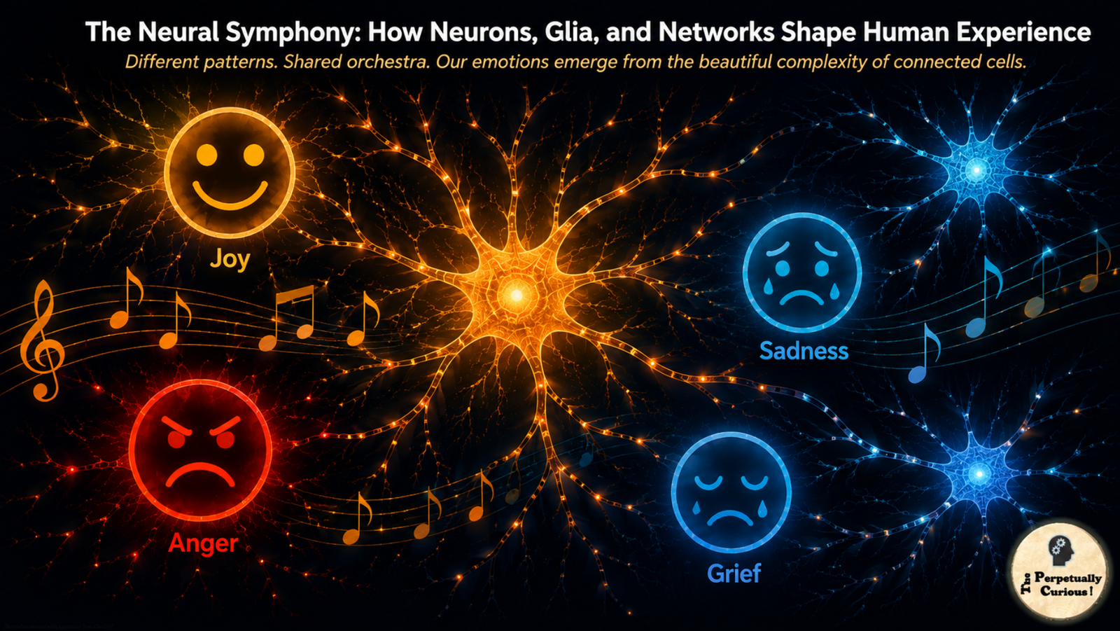
A gentle current moves through the living architecture of the mind, carrying traces of feeling and memory across its quiet distances.
Each signal becomes part of a larger unfolding, shaped by time, rhythm, and the patient work of many unseen hands.
In this still moment, the whole system seems to breathe, holding the fragile beauty of human experience with calm devotion.
⚡ The language of neural communication
Neurons communicate through electricity and chemistry. An action potential can travel along a fast myelinated axon at speeds approaching 394 feet per second (120 meters per second). When that electrical signal reaches a synapse, it can trigger the release of neurotransmitters into a cleft only tens of nanometers wide, often around 20 nanometers at a chemical synapse. These molecules cross the tiny gap and bind to receptors on the next cell, altering the probability that it will respond.
Yet even this familiar neuron-to-neuron picture is incomplete. Astrocytes, star-shaped glial cells, help regulate the chemical environment around synapses. They clear neurotransmitters, buffer ions such as potassium, support neuronal metabolism, and participate in neurovascular coupling, helping match local blood flow and energy supply to neural activity. Astrocytes also show calcium-based signaling that unfolds over slower timescales than electrical spikes. In some experimental contexts they can release signaling molecules such as ATP, although the extent and physiological importance of gliotransmission in the intact human brain remain active areas of research.
Neurotransmitters add another layer of context. Glutamate is the major excitatory neurotransmitter in the mammalian central nervous system, while GABA provides much of its fast inhibition. Dopamine participates in movement, motivation, reward learning, and the assignment of incentive value. Serotonin influences sleep, appetite, mood-related processes, and many other functions through several receptor families. Acetylcholine can sharpen attention in cortex, contribute to memory-related signaling in the hippocampal system, regulate sleep-wake states, and trigger muscle contraction at the neuromuscular junction.
The important point is not that each molecule has one emotional meaning. It is almost the opposite. The effect of a chemical signal depends on where it is released, which receptor receives it, what other signals are present, and what state the larger network is already in.
That dependence on context becomes even clearer when we look at the astonishing diversity of the cells doing the signaling.
🌳 The magnificent diversity of neural architecture
The human brain contains roughly 86 billion neurons, but the word neuron hides enormous diversity. Modern single-cell and single-nucleus methods distinguish thousands of transcriptomic cell types and states across the nervous system, adding molecular detail to older classifications based on shape, location, connectivity, and physiology.
Pyramidal neurons, named for their triangular cell bodies, dominate many excitatory circuits in the cerebral cortex. Excitatory neurons make up roughly four-fifths of cortical neurons, and many of these are pyramidal cells whose long axons connect distant cortical and subcortical regions. Their elaborate dendrites collect input from thousands of synapses, allowing them to integrate signals arriving from many sources.
Interneurons often work on a more local scale. Basket cells can provide powerful inhibition around the cell bodies of neighboring neurons. Chandelier cells target the axon initial segment, a strategically important region for controlling whether a pyramidal neuron fires. Martinotti cells send inhibitory projections toward superficial cortical layers. Stellate cells take several forms across the brain and can serve as compact local processors in cortical and cerebellar circuits.
Purkinje cells of the cerebellum are among the most visually elaborate neurons in the nervous system. Their enormous, flattened dendritic trees receive input from vast numbers of parallel fibers while each Purkinje cell also receives a powerful climbing-fiber input. From this convergence, the cerebellum helps refine movement, timing, prediction, and learning.
The lesson is already emerging: a cell’s identity shapes what it can do, but not what an entire thought or emotion is. Function depends on the network into which that cell is woven.
And neurons are only part of that network.
🌟 The cellular ensemble beyond neurons
For much of neuroscience history, glial cells were treated mainly as support for the supposedly more important neurons. That picture has changed. In the human brain, neurons and non-neuronal cells exist in broadly similar total numbers, although the ratio varies substantially across regions. Glia perform essential roles in metabolism, myelination, immune surveillance, extracellular homeostasis, development, and plasticity.
Astrocytes surround synapses, contact blood vessels, regulate extracellular ions and neurotransmitters, and help distribute metabolic resources. Their signaling is generally slower than the millisecond electrical language of neurons, which means they can influence circuits on different temporal scales.
Microglia are the resident immune cells of the central nervous system. In healthy tissue they continually extend and retract fine processes, surveying the local environment and interacting with synapses and other cells. During development and under particular physiological or pathological conditions, microglia can participate in synaptic remodeling. During injury, infection, chronic stress, or neurodegenerative disease, their states can change dramatically. Those changes may influence neural function, but they should not be imagined as a simple switch between a universally “good” resting state and a universally “bad” activated state.
Oligodendrocytes create myelin around many central nervous system axons. Myelin enables rapid saltatory conduction, allowing some signals to travel at very high speeds along myelinated pathways. Oligodendrocytes also provide metabolic support to axons. Neural activity can influence myelination, but this remodeling occurs over days, weeks, and longer periods rather than moment by moment.
Together, these cells create a brain whose activity unfolds on multiple timescales. Electrical spikes may last milliseconds. Neuromodulatory changes can shape circuits over seconds or minutes. Synaptic plasticity can persist for hours, days, or longer. Myelin and structural remodeling can unfold across weeks or months. Development and aging reshape the same networks across years.
That range of timescales is one reason the brain cannot be understood as a collection of emotional switches.
🎭 The neural cast: key players in the brain’s orchestra
Thousands of neural cell types are still being catalogued, but several broad functional groups help organize the picture.
Projection neurons
Pyramidal neurons: Major excitatory projection cells of the cerebral cortex, connecting local and distant regions through glutamatergic signaling.
Purkinje cells: Large inhibitory neurons of the cerebellar cortex that integrate immense amounts of input and help refine movement, prediction, and timing.
Von Economo neurons: Large, specialized neurons found especially in the anterior cingulate and frontoinsular cortex. Their exact functions remain under investigation, but they have been discussed in relation to rapid integration of socially and internally relevant information.
Magnocellular neurosecretory neurons: Hypothalamic neurons that produce oxytocin or vasopressin and release these hormones into the bloodstream from the posterior pituitary, while related signaling also influences neural circuits.
Interneurons
Basket cells: Often fast-spiking inhibitory neurons that help regulate local timing and synchronization.
Chandelier cells: Specialized cortical interneurons that target the axon initial segments of pyramidal cells.
Stellate cells: Diverse local neurons found in several brain regions, contributing to sensory and computational microcircuits.
Martinotti cells: Inhibitory cortical interneurons that provide feedback regulation across layers of the cortex.
Modulatory neurons
Dopaminergic neurons: Concentrated in several midbrain regions and involved in movement, motivation, learning, and reward-related signaling.
Serotonergic neurons: Located largely in the brainstem raphe nuclei, with widespread projections that influence sleep, appetite, mood-related processing, and other functions.
Noradrenergic neurons: Prominent in the locus coeruleus, helping regulate arousal, vigilance, attention, and adaptive responses to changing conditions.
Cholinergic neurons: Found in the basal forebrain, brainstem, and other regions, contributing to attention, learning, memory, and sleep-wake regulation.
This cast is useful because it shows how specialized individual cells can be. It is also useful because it reveals the limits of cell-by-cell explanation. No entry in the list is a “fear neuron,” “joy neuron,” or “grief neuron.”
To reach emotion, the story must move from parts to patterns.
🎨 From cell types to emotional states
Human emotions are often described through categories such as fear, joy, sadness, anger, grief, love, awe, and contentment. These labels are useful for communication, but they do not map neatly onto one set of cells or one fixed neural program.
Fear can involve threat detection, bodily arousal, memory, attention, action preparation, and conscious interpretation. Grief can involve attachment, memory, yearning, stress physiology, pain-related processing, and social context. Joy can involve reward, surprise, safety, social connection, memory, and bodily expression. Different emotional states can recruit overlapping regions and chemical systems while differing in timing, intensity, context, and the pattern of coordination among them.
This is where a familiar public story begins to break down. If emotions are distributed network states, then calling dopamine a “pleasure chemical” or serotonin a “happiness chemical” cannot capture what those molecules actually do.
🧩 The neurotransmitter paradox: why simple stories persist
Simple chemical stories are attractive because they offer a clean equation: dopamine equals pleasure, serotonin equals happiness, oxytocin equals love. Each contains a fragment of real biology, but each becomes misleading when the fragment is treated as the whole.
Dopamine is deeply involved in reward-related behavior, but reward itself contains separable processes. Dopaminergic signaling contributes strongly to motivation, incentive salience, learning, and prediction-related signals. The hedonic experience of “liking” is not reducible to dopamine alone and involves other systems, including opioid and endocannabinoid mechanisms in particular neural hotspots.
Serotonin is equally difficult to compress into one emotion. Its receptors are distributed across many tissues and brain regions, and different receptor subtypes can have different or even opposing effects. Serotonergic signaling participates in mood-related processes, but also in sleep, appetite, cognition, pain, gastrointestinal function, and many other physiological systems.
Oxytocin is often described as a “love hormone,” yet it is better understood as part of a broader system involved in reproduction, lactation, social salience, bonding, and context-dependent social behavior. Its effects vary with situation, individual history, and the circuits in which it acts.
Myth versus reality
Myth: “A chemical imbalance causes depression.”
Reality: Depression is heterogeneous and involves interacting changes across neural circuits, stress physiology, learning, sleep, immune signaling, genetics, and other biological and psychosocial systems. No single neurotransmitter concentration explains the condition.
Myth: “Dopamine is the pleasure chemical.”
Reality: Dopamine is important for motivation, learning, incentive salience, movement, and several reward-related signals. Pleasure itself is distributed across broader networks and chemical systems.
Myth: “Low serotonin means sadness.”
Reality: Serotonin modulates many functions through multiple receptor types. Emotional state emerges from wider network dynamics rather than from one transmitter level.
Once the simple equations are set aside, a richer question appears: what does turn neural activity into an emotional experience?
🌐 How emotions actually emerge from networks
Think of a city coming alive at rush hour. No single traffic light creates “rush hour.” Cars move through streets, signals change, people make decisions, weather changes behavior, bottlenecks appear, and time of day shapes the entire pattern. The phenomenon exists in the interaction.
Emotion works in a similar way.
Distributed processing across brain regions: Visual or auditory areas identify relevant features of a situation. The hippocampal system contributes memory and context. The amygdala participates in learning about biological significance and threat. The insula helps represent bodily states. Prefrontal regions contribute interpretation and regulation. Motor systems prepare action. The anterior cingulate participates in conflict monitoring, motivation, pain-related processing, and other functions. None of these regions “contains” an emotion by itself.
Chemical context shapes the network: Neuromodulators alter excitability, learning, attention, motivation, and behavioral state across broad populations of cells. Their effect depends on receptor subtype, location, timing, and the activity already present.
Temporal choreography matters: Neural systems do not activate all at once. Rapid sensory and salience signals can be followed by slower contextual evaluation, memory retrieval, bodily feedback, and conscious interpretation. Timing differences from milliseconds to seconds can change how information is integrated, although there is no single timing threshold that flips one emotion into another.
Glial and metabolic context matters too: Astrocytes, oligodendrocytes, microglia, vascular cells, and other non-neuronal partners help maintain the conditions in which circuits operate. Some glial responses are rapid enough to influence ongoing local signaling, while others become especially important during learning, sustained stress, inflammation, injury, development, or disease.
This layered view also explains why the same brain region or neurotransmitter can participate in different experiences. The meaning lies in the pattern.
The idea becomes easier to feel when followed through three familiar emotional landscapes.
🌊 Three emotional sketches: fear, joy, and grief
The following examples are illustrative network sketches, not literal second-by-second recordings of what every human brain does. Emotional experiences vary among individuals and situations, and many of the most detailed cell-level findings come from animal or laboratory studies. The purpose of these sketches is to show how multiple systems can contribute to one experience without assigning an emotion to a single cell type.
😨 Fear: when the world suddenly matters
Imagine hearing footsteps behind you on a dark street.
Auditory pathways rapidly analyze the sound while cortical systems begin identifying its pattern and location. At the same time, circuits involving the amygdala can evaluate biological significance, while the hippocampal system contributes context: where you are, what has happened before, and whether the situation matches a remembered danger. Prefrontal networks add slower interpretation and can either amplify concern or help reappraise it.
If the situation is judged threatening, the hypothalamus and brainstem can recruit autonomic and endocrine responses. The locus coeruleus can increase noradrenergic signaling, heightening vigilance and changing the allocation of attention. Heart rate, breathing, muscle readiness, and sensory priorities shift together.
Astrocytes continue regulating ions, neurotransmitters, and local energy supply as these circuits become active. Microglia remain part of the tissue environment, but an ordinary brief episode of fear should not be described as automatically triggering days of neuroinflammation. Sustained or repeated stress can alter neuroimmune and glial states, which is a different timescale and a different biological claim.
What lingers after the footsteps have passed may be a memory strengthened by attention, arousal, and learning. Fear is therefore not one signal. It is a coordinated state in which perception, memory, bodily response, prediction, and action become temporarily aligned around possible danger.
😊 Joy: when prediction opens into delight
Now imagine turning a corner and unexpectedly seeing a beloved friend.
Visual systems identify the face while memory circuits contribute familiarity and personal significance. Because the encounter is better than expected, dopaminergic circuits can carry reward-related prediction signals and increase motivational salience. Other reward systems contribute hedonic experience, while social and attachment-related networks add the warmth of recognition.
The orbitofrontal cortex, ventral striatum, medial prefrontal regions, amygdala, hippocampal system, and other areas can participate in different aspects of the moment. Facial expression, laughter, approach behavior, and bodily arousal emerge from additional motor and autonomic pathways.
Astrocytes and other glial cells support the local metabolic and chemical environment throughout the activity, but there is no need to imagine a dedicated “joy glia” program switching on. If the encounter becomes part of an enduring relationship or repeated pattern of experience, plastic changes can gradually reshape synapses and network expectations over longer intervals.
Joy, like fear, is therefore a pattern. The difference lies not in one molecule, but in what the brain predicts, remembers, values, and prepares the body to do.
💔 Grief: when attachment meets absence
Grief reveals the same network principle from another direction. A person who has been deeply woven into everyday expectation is suddenly absent, yet memories, habits, attachment signals, and predictions do not disappear at the same moment.
Networks involved in memory and autobiographical thought can repeatedly reactivate representations of the person. Reward and attachment systems that once anticipated contact now encounter a persistent mismatch between expectation and reality. The anterior cingulate, insula, prefrontal cortex, amygdala, hippocampal system, and other regions can contribute to the pain, bodily feeling, recollection, and regulation associated with loss.
For some people, grief also becomes visible through the biology of emotional tears. Crying does not reveal a single “sadness circuit”; it shows how emotion can recruit autonomic, facial, lacrimal, respiratory, and social systems at the same time.
Prolonged severe stress can influence inflammatory signaling and glial physiology, but ordinary grief should not be reduced to microglial activation or a fixed neurotransmitter deficit. Grief is a human process with enormous individual variation, shaped by relationship, memory, culture, circumstances, and time.
Its gradual integration is also a form of plasticity. The person is not erased from memory. Instead, networks learn to hold attachment and absence together.
These three sketches unfold over seconds, minutes, days, and longer periods. That widening timescale leads naturally to another layer of neural organization: the brain itself is rhythmic.
🌙 The temporal brain: neural rhythms across day and night
The brain operates inside nested biological clocks. The suprachiasmatic nucleus of the hypothalamus serves as the principal circadian pacemaker, synchronizing daily rhythms with the light-dark cycle. Yet it is not the body’s only clock. Molecular timekeeping occurs throughout the brain and body, creating the body’s multiple biological clocks that coordinate physiology across different tissues.
During wakefulness, neuromodulatory systems including the noradrenergic locus coeruleus, serotonergic raphe nuclei, cholinergic systems, histaminergic neurons, orexin-producing neurons, and others help maintain alertness and behavioral state. As night approaches under ordinary light-dark conditions, melatonin secretion rises and sleep-promoting networks increasingly oppose wake-promoting systems.
Astrocytes and microglia also show circadian changes in gene expression, metabolism, and signaling. These findings strengthen the view that biological time is a property of cellular communities rather than neurons alone.
Circadian disruption can affect mood, cognition, metabolism, immune function, and sleep. Shift work, irregular light exposure, sleep restriction, and disease can all disturb temporal alignment, although the effects vary among individuals. Neurological disease can also disrupt timing systems, an idea developed further in temporal disruption in Parkinson’s and Alzheimer’s disease.
The daily clock does not stop when consciousness fades. Instead, sleep reveals another form of coordinated neural activity.
💤 The sleeping brain: maintenance, memory, and dreams
Sleep is not neural silence. Brain activity reorganizes into distinctive states that cycle across the night.
During rapid eye movement, or REM, sleep, cortical activity becomes relatively wake-like while vivid dreams are common. Brainstem mechanisms suppress most skeletal muscle activity, reducing the likelihood that dream movements will be acted out. The strange vividness and emotional intensity of dreaming emerge from a changing balance among sensory imagery, memory, emotion, and executive control, a broader story explored in the science of dreaming.
Non-REM sleep contains slower, more synchronized cortical rhythms. Sleep spindles, slow oscillations, and hippocampal sharp-wave ripples participate in the reactivation and consolidation of recently encoded information. Memories are not simply copied from one storage site to another. They are repeatedly reprocessed within distributed systems, and sleep helps determine which traces stabilize, integrate, or fade.
Sleep also changes fluid movement and metabolic clearance in the brain. In landmark mouse studies, sleep and anesthesia were associated with an approximately 60% increase in interstitial space and greater exchange between cerebrospinal and interstitial fluid. This result should not be simplified into the claim that astrocytes themselves shrink by a fixed percentage. Research continues to clarify how sleep state, vascular pulsation, aquaporin-4, and perivascular pathways contribute to glymphatic-like clearance, especially in humans. The wider role of cerebrospinal fluid circulation extends well beyond sleep alone.
Microglia and astrocytes also change their activity across sleep and wake states. What matters is not that one cell type “cleans the brain” while another sleeps, but that sleep changes the physiological environment in which many cellular maintenance processes occur.
Sleep therefore does more than maintain the stage. It helps decide which patterns from waking life will endure.
🔄 The plastic brain: how experience reshapes neural function
Plasticity is the brain’s capacity to change with experience. It includes rapid shifts in synaptic strength, slower structural remodeling, changes in gene expression, altered network connectivity, and activity-dependent changes in myelination.
Long-term potentiation and long-term depression are two well-studied forms of synaptic plasticity. NMDA receptors play important roles in several forms of plasticity, and their activation depends on co-agonists such as glycine or D-serine. The cellular sources and regulation of D-serine vary with brain region and physiological context, with both neuronal and astrocytic contributions discussed in the literature.
Repeated practice can change dendritic spines, synaptic efficacy, and network recruitment. Astrocytes can adjust their interactions with active synapses and help meet metabolic demand. Microglia participate in synaptic remodeling under particular developmental and physiological conditions. Oligodendrocyte precursor cells and oligodendrocytes can respond to neural activity, gradually altering myelination and thereby the timing and reliability of communication across circuits.
Experience can also influence gene expression and epigenetic regulation in neurons and glia. These changes do not mean that every thought permanently rewrites the brain. They mean that repeated, salient, or biologically important experience can leave molecular and structural traces that alter how future signals are processed.
Development provides the clearest example. Early in life, neurons and synapses are produced in abundance and then refined through activity, competition, stabilization, and pruning. Astrocytes promote synapse formation and regulate the extracellular environment. Microglia participate in developmental remodeling. Experience interacts with this biology during sensitive periods, helping shape circuits that later support sensation, movement, cognition, and emotion.
Even in adulthood, memory systems remain dynamic. The brief separation between familiarity and recollection that appears in déjà vu offers one small window into how distributed memory processes can momentarily fall out of alignment.
Because experience changes circuits, other people become part of the environment that changes them. That brings the cellular story into the social world.
🤝 The social brain: action observation, resonance, and empathy
Mirror neurons were first identified in macaque monkeys in the 1990s. These cells respond both when an animal performs certain actions and when it observes similar actions performed by another.
Humans also show a well-supported action-observation network involving premotor, inferior frontal, and parietal regions, with activity overlapping between performing and observing actions. Some invasive human recordings have identified neurons with mirror-like response properties, but much of the human evidence comes from imaging, electrophysiology, and stimulation methods that measure populations rather than single cells.
For that reason, it is safer to speak of a human action-observation or putative mirror system than to claim that individual mirror neurons directly create empathy. Empathy itself includes several partially separable processes, from sharing another person’s affective state to understanding perspective and choosing a social response.
Observing another person in pain can recruit parts of the anterior insula and anterior cingulate cortex that also participate in first-person pain-related processing, although the patterns are not identical. The temporoparietal junction, medial prefrontal cortex, amygdala, memory systems, and other regions contribute to perspective-taking and social inference. Oxytocin and other neuromodulators can influence social salience in context-dependent ways.
The social brain is therefore another ensemble. Action resonance may contribute to understanding others, but empathy cannot be assigned to one neuron class any more than joy or grief can.
These social circuits remain plastic across decades, which leads from momentary connection to the longer biological arc of aging.
🌅 The aging brain: resilience and adaptation
Aging changes the brain, but it does not simply empty it of neurons. Many cortical neuron populations are relatively well preserved during normal aging, while particular cell types and regions can show greater vulnerability. Changes in synapses, dendrites, white matter, vascular function, neurotransmission, inflammation, and network efficiency all contribute to age-related cognitive differences.
Astrocytes and microglia change with age as well. Their gene-expression patterns, morphology, inflammatory responsiveness, and homeostatic functions can shift in region-specific ways. Oligodendrocytes and their precursor cells continue participating in myelin maintenance, although white matter integrity can decline with age.
One long-running question has been whether the adult human hippocampus continues to generate new neurons. Earlier studies produced conflicting results. In February 2026, a large multiomic study of the human hippocampus reported neural stem cells, neuroblasts, and immature granule neurons across adulthood and aging, strengthening the evidence that adult hippocampal neurogenesis persists. Important questions remain about its rate, individual variability, regulation, and contribution to human cognition.
Exercise provides another example of plasticity without requiring a single-cell explanation. In an influential year-long randomized trial of older adults, aerobic exercise increased anterior hippocampal volume by about 2%, while the control group showed decline. Later research has continued to investigate how vascular, metabolic, trophic, inflammatory, and neural mechanisms contribute to exercise-related brain effects.
Aging can also bring strengths. Crystallized knowledge, vocabulary, expertise, and some forms of emotional regulation can remain stable or improve across much of adulthood. Functional imaging has shown that older adults sometimes recruit broader or more bilateral networks during cognitive tasks, including patterns described by the HAROLD model of reduced hemispheric asymmetry. Such compensation is not universal, but it illustrates the brain’s ability to reorganize rather than simply surrender function.
Resilience, however, is not invulnerability. The same systems that adapt across a lifetime can also become vulnerable to disease.
🏥 When networks falter
Neurological and psychiatric conditions reveal how misleading single-cause stories can be.
Major depressive disorder is heterogeneous. Group-level studies have identified differences in connectivity, stress regulation, sleep, inflammatory signaling, and brain structure, including modest average reductions in hippocampal volume in some meta-analyses. Astrocytic and microglial alterations have also been reported in animal models, postmortem studies, and some human imaging work. These findings support a role for glial biology in subsets of depression, but they do not establish one universal “glial cause” of depressive illness.
Parkinson’s disease offers a clearer example of selective neuronal vulnerability. Degeneration of dopamine-producing neurons in the substantia nigra pars compacta is central to the classic motor syndrome. Yet Parkinson’s disease extends beyond dopamine. Protein handling, mitochondrial stress, autonomic systems, sleep, inflammation, and distributed neural networks can all become involved. The broader relationship between altered proteins and selectively vulnerable cells is explored through protein misfolding in neurodegenerative disease.
Alzheimer’s disease likewise cannot be reduced to the death of one cell type or the presence of one protein deposit. Amyloid, tau, synaptic dysfunction, glial responses, vascular factors, genetics, and network vulnerability interact across years.
These conditions demonstrate the same principle as healthy emotion and cognition: the brain’s properties arise through interactions among cell identity, molecular state, connectivity, timing, and environment.
That complexity is precisely why modern neuroscience is moving beyond simply naming brain parts.
🔬 The future of neural understanding
Modern neuroscience can now ask questions that were almost unimaginable a few decades ago.
Optogenetics allows researchers to control genetically defined cell populations with light in experimental animals, providing precise tests of circuit function. Chemogenetic and molecular tools offer additional ways to manipulate neuronal and glial populations. Single-cell and single-nucleus sequencing reveal molecular diversity that older anatomical classifications could not see.
Large-scale electrophysiology records from many neurons simultaneously, while advanced microscopy tracks activity and structure across living tissue. Machine learning can help identify patterns in these enormous datasets and, in controlled settings, decode aspects of visual input, intended movement, or other neural variables.
Brain organoids add another tool. These three-dimensional cultures derived from stem cells can model selected features of human neural development and disease. They can contain multiple neural and glial cell types and can develop organized regional features, but they are not miniature replicas of complete human brains. Their value lies in creating experimentally accessible models of particular developmental processes.
As measurement improves, the deepest questions become sharper rather than simpler. How do cellular events become perception? How do distributed networks maintain a unified sense of self? How does activity become conscious experience? These questions reach beyond any one neuron, synapse, or theory.
The closer neuroscience looks, the less the brain resembles a soloist and the more clearly it resembles an ensemble.
🎵 The infinite symphony continues
As you finish reading these words, billions of neurons and tens of billions of glial cells continue their ceaseless activity. Electrical impulses cross membranes. Chemical signals bind to receptors. Astrocytes regulate local environments. Oligodendrocytes maintain the insulation of long axons. Microglia survey the tissue around them. Blood vessels adjust to changing metabolic demand.
Learning about the brain can itself change the brain, but not because one paragraph instantly builds a permanent new circuit. Attention and learning can alter synaptic activity within moments, influence gene expression over longer intervals, and, when experience is repeated or important, contribute to lasting changes in connectivity and network function.
That may be the deeper wonder of neuroscience. The organ trying to understand itself is not fixed while it performs the investigation. It is living tissue, shaped by development, memory, sleep, relationships, illness, recovery, and time.
The next time joy rises unexpectedly, fear sharpens the world, or memory carries someone absent into the present, there is no need to search for a single responsible cell. Beneath awareness, countless processes are converging across different places and timescales. The experience feels singular because the orchestra plays as one.
In that unity, complexity does not diminish the mystery. It gives the mystery structure.
Pass this article along to someone curious and let the learning travel.
💡 Did You Know
🔥 The brain’s energy appetite: The adult human brain accounts for only about 2% of body mass but uses roughly 20% of the body’s resting energy expenditure. Much of that energy supports electrical signaling, ion gradients, synaptic transmission, maintenance, and the cellular systems that keep neural tissue functioning.
⏳ Some neurons may last a lifetime: Many neurons in the cerebral cortex are generated before birth and can persist for decades. Their longevity means that the same cells can participate in changing networks across childhood, adulthood, and old age even as their synaptic connections are continually remodeled.
🚂 A single cell can span an extraordinary distance: Some motor neurons extend axons from the spinal cord to muscles in the foot, reaching more than 3 feet (about 1 meter) in tall adults. The cell must transport proteins, organelles, and signals across this remarkable internal distance while maintaining communication with its target.
🐙 Intelligence does not require the vertebrate layout: Octopuses have roughly half a billion neurons, with a large proportion distributed through their arms. Their nervous system shows how substantial local processing can occur outside a centralized vertebrate-style brain, one of the features that makes octopus intelligence such an intriguing comparison with our own neural architecture.
🌌 The brain contains an immense number of synapses: Estimates vary by region and method, but the human brain is commonly thought to contain on the order of 100 trillion synapses. The number matters less than what it represents: cognition emerges from relationships among cells, not from the cells in isolation.
What is the main difference between neurons and other cells in the body?
Neurons are specialized for rapid electrical signaling and long-distance communication. Many have dendrites for receiving input, axons for sending signals, and synapses for communicating with other cells. Mature neurons in most brain regions generally do not divide, although the nervous system also contains progenitor cells and limited neurogenic niches.
How many types of neurons exist in the human brain?
There is no single final number. Modern transcriptomic methods reveal thousands of molecularly distinct cell types and states, while anatomical and physiological classifications divide neurons in other ways. The number depends partly on what scientists mean by a “type.”
Can a specific emotion be traced to one neuron or one brain region?
No. Particular neurons and regions can be important for particular components of emotion, but a subjective emotional state emerges from distributed activity involving perception, memory, bodily regulation, motivation, learning, and context.
Do glial cells contribute to emotion?
Glial cells contribute to the physiological environment in which emotional circuits function. Astrocytes regulate neurotransmitters, ions, metabolism, and synaptic conditions; oligodendrocytes influence signal timing through myelin; and microglia interact with synapses and immune pathways. Evidence also links glial changes with chronic stress and mood disorders. What remains uncertain is how specific glial events map onto ordinary moment-to-moment human emotions.
Do adults grow new neurons?
Current evidence supports the persistence of at least some hippocampal neurogenesis in adults. A major 2026 multiomic study strengthened this conclusion by identifying neural stem cells, neuroblasts, and immature granule neurons across adult human samples. The rate of new-neuron production, how much it varies among people, and its functional importance in human memory and mood remain active research questions.
How do neurons communicate with one another?
Most neural communication occurs at chemical synapses, where an electrical signal in one neuron triggers neurotransmitter release and changes the activity of another cell. Some neurons also communicate through electrical synapses formed by gap junctions. Both forms of signaling operate within a larger cellular environment shaped by glia, extracellular ions, blood flow, and neuromodulators.
What happens to the brain during sleep?
Neural activity changes rather than stops. Different sleep stages contain characteristic rhythms, including slow oscillations, sleep spindles, and REM activity. Sleep supports memory processing, metabolic regulation, and other maintenance functions. Fluid exchange through perivascular pathways also changes with sleep state, although the exact mechanisms and their importance in humans continue to be investigated.
Are mirror neurons responsible for empathy?
Not by themselves. Humans have action-observation networks that overlap when actions are performed and observed, and some neurons with mirror-like properties have been recorded directly. Empathy, however, also involves emotional, memory, perspective-taking, and regulatory systems. Mirror-like processes may contribute to social understanding, but they do not provide a complete explanation.
Can damaged neurons regenerate?
Peripheral nerves can regrow axons to some extent after injury, often at rates on the order of 0.04 inches (about 1 millimeter) per day under favorable conditions. Regeneration in the adult brain and spinal cord is far more limited because of both intrinsic neuronal constraints and the surrounding tissue environment.
What happens in the brain when we learn something new?
Learning changes patterns of neural activity and can alter synaptic strength within minutes. With consolidation and repetition, changes in gene expression, dendritic structure, network recruitment, and sometimes myelination can help stabilize new skills or memories. The timescale depends on the kind of learning, and not every new fact produces a permanent structural change.
🌸 Sharing the wonder
We kindly invite you to share and spread the word about the marvels within the mind. If this exploration offered a new way to think about neurons, glia, emotion, and the networks that hold them together, pass it along to someone whose curiosity might follow the next signal.
A single question can travel farther than a single neuron ever could.
“The Neural Symphony: How Neurons, Glia, and Networks Shape Human Experience.” The Perpetually Curious!, August 2026.
https://www.theperpetuallycurious.org/articles/how-the-brain-creates-emotions/Continue Exploring
For the curious mind: want to explore this theme through a few questions? Visit the Classic Quiz Studio, nestled under Explorations opens in a new tab ,and see where a few well-placed questions lead: Biology & Health Quizzesopens in a new tab
For what comes next
Site Updates gathers occasional notes, new additions, and ways to keep in touch. RSS brings new publications to your feed reader in chronological order.
Choose the path that speaks to you, and let the next curiosity find you wherever your wondering takes you.