

At first glance, a protein is only a chain of amino acids. Inside the cellular metropolis of a neuron, that chain must bend, twist, and settle into a three-dimensional form precise enough to carry out its task. It is a kind of molecular origami repeated continuously across the nervous system, shaping receptors, enzymes, scaffolds, transport machinery, and countless other parts of cellular life.
The cell does not leave this choreography to chance. Molecular chaperones assist proteins as they fold and refold, while quality-control systems such as the ubiquitin-proteasome pathway and autophagy help remove proteins that cannot be repaired. Most folding errors never become disease. Trouble begins when particular proteins adopt harmful conformations that persist, seed further misfolding, interfere with cellular machinery, or accumulate faster than the cell can contain or clear them.
That distinction matters because neurodegeneration is rarely the story of one bad molecule. It is the story of abnormal protein biology meeting a vulnerable cell over time. Alzheimer’s disease, Parkinson’s disease, and amyotrophic lateral sclerosis each reveal a different version of that encounter, and together they show why protein shape is only the beginning of the story.
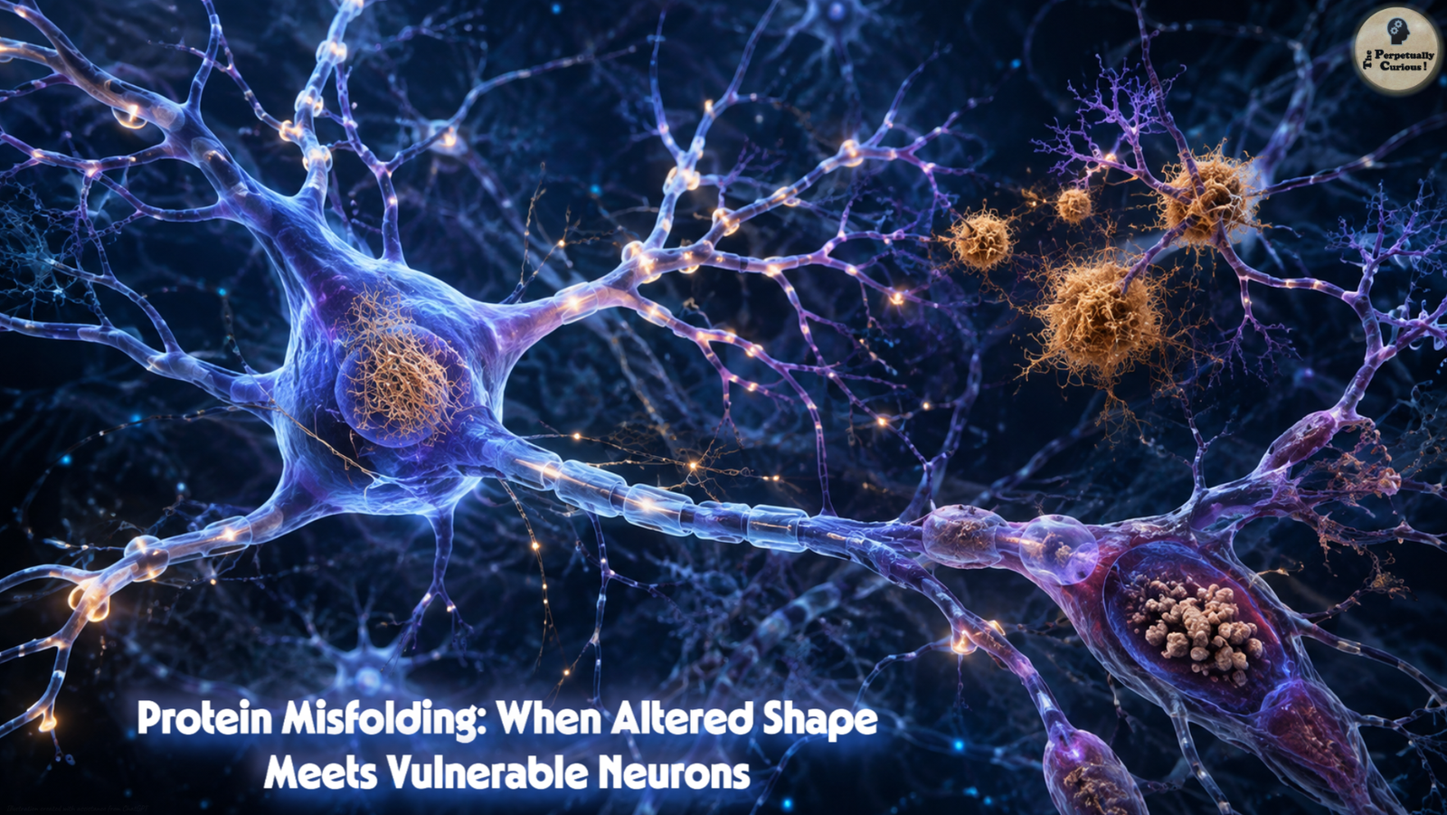
A single line of thought drifts through the living architecture of the brain, shaped by forces both fragile and immense.
Proteins rise and fall in their quiet choreography, carrying the memory of form across the years.
In their delicate balance, the story of our humanity continues its patient unfolding.
🌀 The misfolding cascade
Some disease-associated proteins can adopt conformations that encourage additional copies of the same protein to misfold or aggregate. This seeding behavior can allow abnormal protein states to amplify, while the resulting oligomers, fibrils, inclusions, and cellular stress responses disturb different parts of neuronal function. The details vary by disease, but the recurring theme is a growing mismatch between what the cell must manage and what its quality-control systems can safely contain.
In Alzheimer’s disease, amyloid-β peptides can assemble into soluble oligomers and fibrils that accumulate as extracellular plaques. Smaller soluble species and plaque-associated processes can disrupt synaptic signaling, alter cellular metabolism, and contribute to inflammatory responses involving microglia. Inside neurons, tau follows a different path. Normal tau helps support microtubules, which are part of the transport system that moves materials through long neuronal processes. When tau becomes abnormally modified and aggregates, loss of normal tau function together with toxic tau species can impair transport and synaptic function. Soluble tau assemblies may contribute to toxicity before mature neurofibrillary tangles become prominent.
Tau pathology often appears early in transentorhinal and entorhinal regions before involving wider cortical networks. Those regions are deeply connected with memory systems, helping to place early memory impairment within a broader anatomical pattern. The relationship is not a simple one-to-one explanation of symptom order, but it shows how a molecular change can become a network-level disease.
Parkinson’s disease tells a related story through alpha-synuclein. Misfolded and aggregated alpha-synuclein is a major component of Lewy pathology, which appears across several nervous-system regions as the disease develops. In many cases, pathology is found early in olfactory or lower-brainstem regions in patterns consistent with Braak-type staging, although Parkinson’s disease does not follow one universal anatomical sequence. Hyposmia and REM sleep behavior disorder can precede the characteristic motor syndrome in some people.
The motor features of Parkinson’s disease are closely tied to the loss of dopamine-producing neurons in the substantia nigra pars compacta. These neurons carry unusual metabolic and structural burdens, and their degeneration reduces dopamine signaling to movement-related circuits. Tremor, rigidity, and slowed movement can emerge as that system loses resilience, while cognitive changes may develop later in some people as additional neural networks become involved.
Amyotrophic lateral sclerosis, or ALS, reveals the same broad problem through a different cellular landscape and often a much faster clinical course. TDP-43 pathology is present in most ALS cases, while misfolded mutant SOD1 is central to SOD1-associated ALS. These proteins do not represent one universal ALS mechanism, but they illustrate how disturbed protein handling can intersect with RNA processing, axonal transport, mitochondrial stress, and other cellular systems. Motor neurons progressively fail, producing weakness and paralysis. Median survival is often about 2 to 5 years after symptom onset, although individual trajectories vary widely.
The three diseases therefore share a molecular theme without becoming versions of the same disorder. The proteins differ, the cells differ, the networks differ, and the tempo differs. That leads naturally to a harder question: if abnormal proteins can be present across broad areas of the nervous system, why do some neurons fail before others?
🧩 The architecture of vulnerability
A neuron is not merely a generic cell carrying electrical signals. Different neuron classes have different shapes, firing patterns, energy demands, chemical environments, axonal lengths, and relationships with surrounding glial cells. Those differences help explain why the same broad category of cellular stress can produce sharply selective patterns of degeneration.
Motor neurons illustrate the problem of scale. Some extend axons close to 3 feet (1 meter) from the spinal cord to distant muscles. Proteins, organelles, nutrients, and molecular signals must be transported across that extraordinary distance while the neuron maintains a large cell body, an extensive axon, and thousands of synaptic relationships. When proteostasis, energy production, or axonal transport becomes strained, the logistical burden of maintaining such a cell can become especially consequential.
Dopaminergic neurons of the substantia nigra face a different combination of pressures. Dopamine chemistry can generate reactive byproducts, while autonomous electrical activity and calcium handling contribute to metabolic demand. Many of these neurons also maintain enormous, highly branched axonal arbors. Calcium entry through L-type channels is one contributor to this burden, but it is not a single explanation for Parkinson’s disease. Oxidative stress, mitochondrial function, dopamine metabolism, proteostasis, aging, genetics, and network architecture intersect in determining vulnerability.
The contrast becomes especially revealing in ALS. Oculomotor neurons that control eye movements often show relative resistance compared with many spinal motor neurons, even as the disease advances. That resistance is not absolute, and no single protective mechanism explains it. Still, comparing vulnerable and relatively resistant cells gives researchers a way to look for protective molecular programs rather than studying degeneration alone.
That uneven vulnerability also reflects neuronal diversity, because different neuron classes and their supporting glial environments do not carry the same structural, metabolic, or proteostatic burdens. Protein identity, then, is only half the map. The other half is the cell and circuit in which that protein appears.
🎯 How vulnerability becomes a disease pattern
Distinct neurodegenerative diseases emerge from the intersection of several layers: which protein is altered, which cellular systems are stressed, which neuron classes are most vulnerable, and how those neurons are connected to one another. Selective vulnerability therefore does not mean that one protein simply targets one cell type. It means that particular molecular and cellular conditions make some parts of the nervous system less able to absorb a growing burden.
In Alzheimer’s disease, tau pathology tends to progress through recognizable regional patterns and is associated with connected memory networks. In Parkinson’s disease, alpha-synuclein pathology can involve multiple neural systems while the loss of substantia nigra dopamine neurons remains central to the classic motor syndrome. In ALS, disease-related proteins may be expressed widely, yet the motor system bears the defining clinical burden.
Cell-to-cell propagation may also contribute to the way some protein pathologies expand through connected tissue. Researchers sometimes describe this as prion-like behavior because a misfolded protein can seed additional misfolding within a nervous system. The term describes a mechanistic resemblance, not ordinary person-to-person contagion.
This framework also marks an important boundary. Not every neurological disease is primarily a proteinopathy. Multiple sclerosis, for example, is driven mainly by immune-mediated inflammation and demyelination rather than by the accumulation of a misfolded disease protein.
Once protein state, cellular vulnerability, and network context are viewed together, the treatment question changes. The challenge is no longer simply how to remove one abnormal protein. It becomes a broader question of where the disease chain can be interrupted without losing sight of the neurons that must survive.
🔬 From understanding to treatment
Many established therapies still work mainly by improving function or relieving symptoms. Levodopa is converted to dopamine and helps restore dopamine signaling in Parkinson’s disease. Cholinesterase inhibitors and memantine act through different neurotransmitter systems to support cognition or daily function in Alzheimer’s disease. These treatments remain important even when they do not remove the underlying molecular pathology.
Disease-modifying therapies now add another layer. Lecanemab and donanemab are amyloid-directed antibodies approved for selected people with early symptomatic Alzheimer’s disease. By reducing amyloid pathology, they can slow clinical decline at the group level, but they do not halt or reverse the disease. Both can cause amyloid-related imaging abnormalities, or ARIA, which may involve brain edema or small hemorrhagic changes, so their use is accompanied by clinical and imaging surveillance.
A different strategy is already visible in a defined form of ALS. Tofersen is an antisense oligonucleotide approved under the accelerated approval pathway for adults with ALS associated with an SOD1 mutation. It targets SOD1 messenger RNA and lowers production of SOD1 protein. Its approval was based on reduction of neurofilament light, a biomarker of axonal injury that is considered reasonably likely to predict clinical benefit, while confirmatory evidence of clinical benefit continues to be studied.
Beyond these approved examples, researchers are testing ways to strengthen the cell’s own protein-quality-control network. Molecular chaperones can assist folding and refolding, limit aggregation, or help route proteins toward degradation. The proteasome and autophagy-lysosome systems remove different classes of damaged proteins and cellular material. Rather than behaving like one repair crew, these pathways form a distributed proteostasis network whose capacity can change with age, stress, and disease.
The architecture of resilience is becoming a therapeutic question as well. If relatively resistant neurons possess molecular programs that help them handle calcium, oxidative stress, misfolded proteins, or long-distance transport, those programs may reveal candidates for neuroprotection. The aim is not to turn every neuron into another cell type, but to understand which protective features can be strengthened without disrupting normal function.
The therapeutic picture therefore returns to the same symmetry: the protein matters, the neuron matters, and timing matters. If harmful biology can be detected earlier, intervention may have a larger window before extensive neuronal loss has occurred.
🌅 The horizon of hope
Biomarkers are beginning to make some parts of neurodegenerative disease visible before the full clinical picture is established. In research on autosomal-dominant Alzheimer’s disease, certain amyloid and tau changes can appear many years before the expected onset of symptoms, although different markers change on different timelines. PET imaging can reveal amyloid or tau pathology in the living brain, while cerebrospinal fluid biomarkers can show characteristic shifts in amyloid and phosphorylated tau.
Blood-based testing is also moving rapidly. In 2025, the U.S. Food and Drug Administration cleared its first blood test to aid the evaluation of amyloid pathology associated with Alzheimer’s disease, using a plasma pTau217 to amyloid-β 1-42 ratio. The test is intended to support evaluation of symptomatic adults in specialized care, not to screen the general population or provide a stand-alone diagnosis. That distinction is important because a biomarker is a measurement of disease biology, not a complete forecast of one person’s future.
Earlier detection does not make neurodegenerative disease simple. It does, however, change the questions researchers can ask. Can treatment begin before a vulnerable network has lost too many cells? Can molecular subtypes help identify which therapy is most relevant? Can the protective biology of resistant neurons be understood well enough to preserve function longer? These questions move the field from watching late consequences toward testing earlier points of intervention.
The deeper lesson is not that every neurodegenerative disease shares one culprit. It is that protein shape, protein clearance, cellular identity, and network context interact across time. A protein only nanometers across can become one thread in a process that alters movement, memory, speech, and independence. Understanding that chain does not make these diseases easy to solve, but it makes the questions sharper, and sometimes that is where progress begins.
🧬 The molecular cascade at a glance
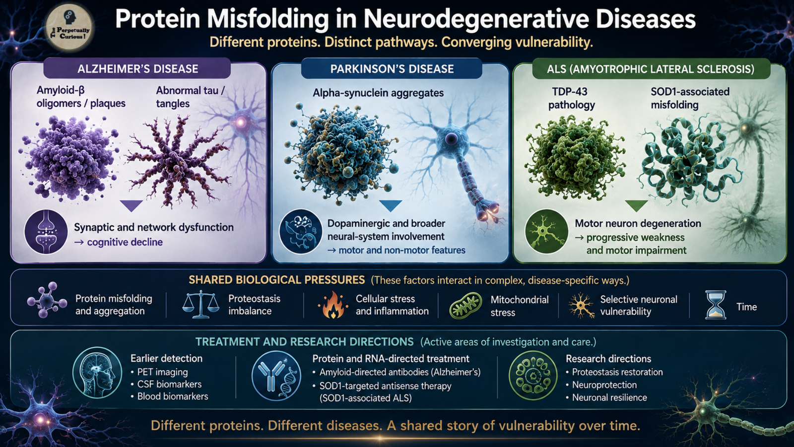
From protein misfolding to selective neuronal vulnerability, this visual synthesis follows three disease pathways: amyloid-β and tau in Alzheimer’s disease, alpha-synuclein in Parkinson’s disease, and TDP-43 or SOD1-associated pathology in ALS. The molecular routes differ, but each shows how altered protein states can intersect with cellular burden and vulnerable neural networks.
Pass this article along to someone curious and let the learning travel.
Did You Know
🧪 Some therapies against protein misfolding work by stabilizing a protein rather than removing an aggregate. Tafamidis binds transthyretin and helps stabilize its normal tetrameric form, slowing the dissociation step that can lead to transthyretin amyloid formation. Although transthyretin amyloidosis is a different disease family from the brain disorders discussed above, the strategy shows that preventing a harmful conformational transition can itself be therapeutically useful.
🩸 Neurofilament light is a structural protein found in axons. When axons are injured, neurofilament light can rise in cerebrospinal fluid and blood across several neurological conditions. That makes it useful as a marker of neuronal or axonal injury, but not as a diagnosis of one specific neurodegenerative disease.
Why do different neurons show different vulnerability?
Neurons differ in axonal length, branching, firing pattern, calcium handling, energy demand, neurotransmitter chemistry, protein-quality-control capacity, and relationships with surrounding glial cells. These features create different biological burdens. A stress that one neuron can absorb may become more consequential in another cell that already operates close to its metabolic or transport limits.
Are plaques, tangles, and Lewy bodies always the most harmful protein forms?
Not necessarily. Large deposits are important pathological hallmarks, but smaller soluble oligomers and other intermediate conformations can also disrupt synapses, membranes, protein interactions, and cellular signaling. The most damaging species may differ by protein, disease stage, and cellular context, which is one reason visible aggregates do not tell the entire mechanistic story.
How do misfolded proteins damage neurons?
Several mechanisms can contribute. Misfolded or aggregated proteins may interfere with normal protein interactions, stress the proteasome and autophagy systems, disturb membranes or calcium balance, impair axonal transport, alter mitochondrial function, and provoke inflammatory responses. Some disease-associated proteins can also seed additional misfolding or move between connected cells. No single mechanism explains every proteinopathy.
Can protein-related changes be detected before symptoms appear?
In some diseases and research settings, yes. Amyloid and tau changes associated with Alzheimer’s disease can sometimes be detected by cerebrospinal fluid analysis, PET imaging, or blood-based biomarkers before dementia is established, and some biomarker changes can precede expected symptoms by many years in inherited Alzheimer’s disease. Detection does not by itself establish when symptoms will begin or how rapidly disease will progress.
How can inherited mutations contribute to these diseases?
Inherited variants can influence neurodegeneration in different ways. Some alter the sequence or amount of a disease-associated protein, while others affect protein processing, RNA biology, trafficking, or clearance. SOD1 mutations can cause a defined form of ALS, while C9orf72 repeat expansions are another major genetic cause within the ALS-frontotemporal dementia spectrum. Rare mutations in genes such as APP, PSEN1, and PSEN2 can cause familial early-onset Alzheimer’s disease. Other genetic variants modify risk without making disease inevitable.
What influences the speed of progression?
Progression is shaped by many interacting factors, including the disease subtype, which neurons and networks are involved, age at onset, genetic background, cellular stress responses, inflammation, and the ability of neural systems to compensate for damage. These influences differ across diseases and among individuals, so one timeline cannot describe every case.
Are Alzheimer’s disease, Parkinson’s disease, and ALS contagious?
No. These diseases do not spread between people through ordinary contact. When researchers use the term prion-like to describe tau, alpha-synuclein, or other disease-associated proteins, they are referring to the ability of a misfolded protein state to seed or propagate within cells and neural networks. That is different from person-to-person transmission. True prion diseases are a separate group of disorders with distinct biology.
✨ Share the wonder
At the scale of a protein, shape can seem almost invisible. Across the lifetime of a neuron, it can become one thread in a much larger biological story. If this journey through molecular landscapes sparked curiosity, share it with someone who enjoys seeing how the smallest structures can illuminate the largest questions of movement, memory, and cellular life.
🧬 Continue exploring this theme
More articles on living systems, human health, cellular processes, and the patterns that shape life are gathered under Biology & Health.
🧠 A gentle note: This exploration of neurodegenerative disease is intended for educational understanding and does not provide individual medical guidance.
“When Proteins Forget Their Shape: Protein Misfolding in Neurodegenerative Diseases.” The Perpetually Curious!, August 2026.
https://www.theperpetuallycurious.org/articles/protein-misfolding-neurodegeneration/Continue Exploring
For the curious mind: want to explore this theme through a few questions? Visit the Classic Quiz Studio, nestled under Explorations opens in a new tab ,and see where a few well-placed questions lead: Biology & Health Quizzesopens in a new tab
For what comes next
Site Updates gathers occasional notes, new additions, and ways to keep in touch. RSS brings new publications to your feed reader in chronological order.
Choose the path that speaks to you, and let the next curiosity find you wherever your wondering takes you.